Japanese
English
- 有料閲覧
- Abstract 文献概要
- 1ページ目 Look Inside
I.はじめに
外傷性一次性脳幹損傷のCT診断においてCT固有の頭蓋底からのartifactsおよび分解能の限界が大きな問題点であった.MRIの登場はこれを克服しうる画期的な出来事といえるが,普及率の点から現在も脳幹損傷の画像診断はCTを中心に行われている15).その際,中脳実質内の異常所見の有無にかかわらず,迂回槽内に孤立した血腫の存在を認めることがある.この迂回槽出血は脳幹表面を走行する血管の破綻によって生じたと考えられ,その部位に損傷が加わったことを間接的に示す所見として大変興味深いが,診断価値に関して検討はほとんどなされていない.
われわれはCT上の孤立した迂回槽出血を呈した4症例を経験し,これらについてMRIによる脳幹実質の損傷の有無を検討し興味ある知見を得たので報告する.
Traumatic hemorrhage in the ambient cistern isthought to be an indirect indication of brain stem injury. In many cases such brain stem lesions cannot be clearly demonstrated by conventional CT scans. Magnetic reso-nance imaging (MRI) provides a more sophisticated dis-play of the brain stem with improved contrast resolution of structures not appreciated on CT.
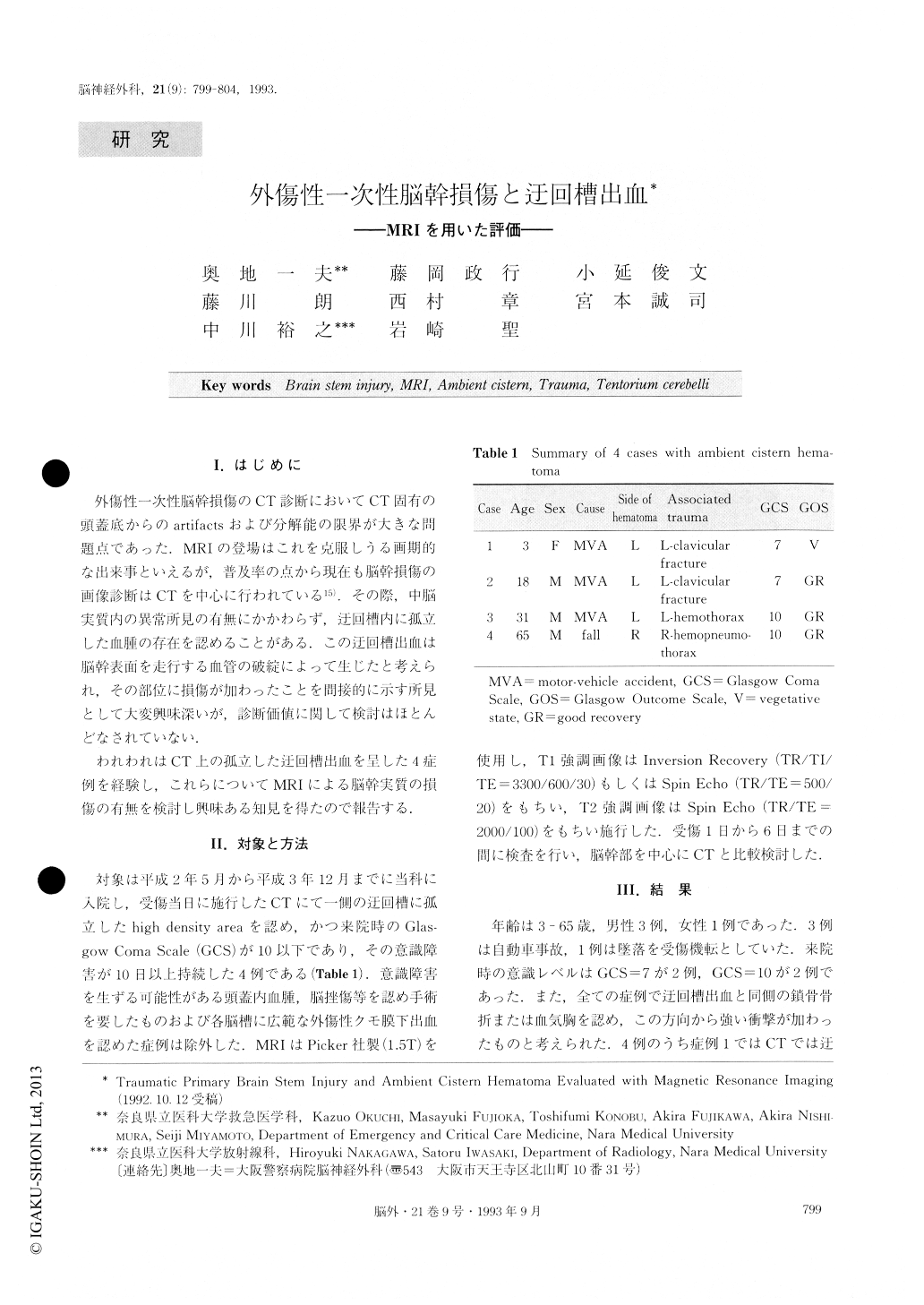
We present four patients with traumatic ambient cis-tern hematoma on CT. They showed consciousness dis-turbance at the initial neurological examination and a Glasgow Coma Scale (GCS) of 7-10. All patients had hemothorax or clavicular fracture ipsilateral to the ambient cistern hematoma that suggested a severe mechanical force from the same direction. Axial, coronal and sagittal MRI scans were obtained with a super-conductive 1.5 T unit (Picker) within 6 days after trauma. Two standard pulse sequences were used ; (1) Spin-echo (TR/TE = 500/20) or Inversion Recovery (TR/TI/TE = 3300/600/30) to obtain Tl-weighted in-formation and (2) Spin-echo (TR/TE=2000/100) to obtain T2-weighted information.
In case 1 (3-year-old girl) the hematoma which was thought to be located in the ambient cistern on CT was found to be present in the subpial region in the tegmen-tum on MRI. On T2 weighted image, a high signal in-tensity area was seen in the perifocal area. This area was demonstrated as a low density area on CT. This patient has remained in a persistent vegetative state 6 months af-ter trauma. In case 3 (31-year-old man) CT demonstrated no abnormal findings in the brain stem. MRI demon-strated a high intensity area in the right cerebral peduncle and left tegmentum. The nonhemorrhagic lesions were diagnosed as local brain edema. In case 2 (18-year-old man) and case 4 (65-year-old man) no abnormal brain stem findings were noted by either MRI or CT. All pa-tients except for case 1 became alert from 1 week to 1 month after trauma.
The resolution of conventional CT is not sufficient to detect lesions of the brain stem. MRI is of great value in the detection of small brain stem injury and for making a prognosis. Isolated ambient cistern hematoma on CT is a finding indicative of mild brain stem injury.
Copyright © 1993, Igaku-Shoin Ltd. All rights reserved.