Japanese
English
- 有料閲覧
PPV購入案内
1,320円 (1,200円+税10%) こちらは、843ページから846ページの文献です。購入には医書.jp本会員登録が必要です。
会員登録完了後に改めて上記「購入サイトへ」を選択してください。
または「購入サイトへ」選択後に「購入ログイン」、「新規会員登録」の順に進んでください。
会員登録について詳しくはをご確認ください。
- Abstract 文献概要
- 1ページ目 Look Inside
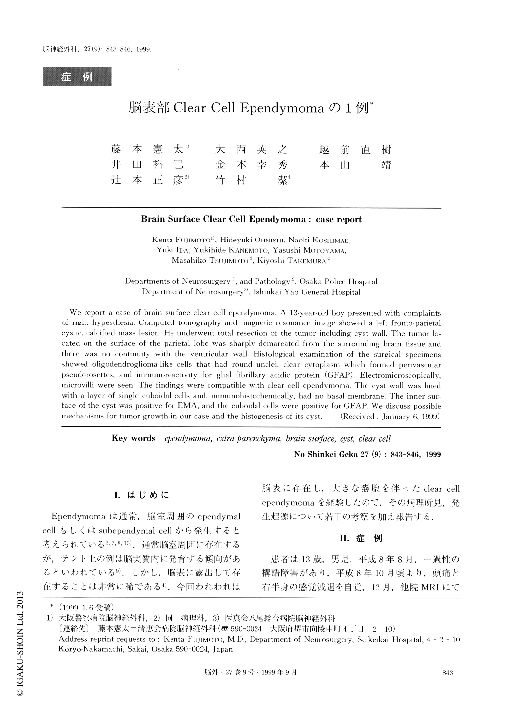
I.はじめに
Ependymomaは通常,脳室周囲のependymalcellもしくはsubependymal cellから発生すると考えられている2,7,8,10).通常脳室周囲に存在するが,テント上の例は脳実質内に発育する傾向があるといわれている9).しかし,脳表に露出して存在することは非常に稀である4).今回われわれは脳表に存在し,大きな嚢胞を伴ったclear cellependymomaを経験したので,その病理所見,発生起源について若干の考察を加え報告する.
We report a case of brain surface clear cell ependymoma. A 13-year-old boy presented with complaintsof right hypesthesia. Computed tomography and magnetic resonance image showed a left fronto-parietalcystic, calcified mass lesion. He underwent total resection of the tumor including cyst wall. The tumor lo-cated on the surface of the parietal lobe was sharply demarcated from the surrounding brain tissue andthere was no continuity with the ventricular wall. Histological examination of the surgical specimensshowed oligoclendroglioma-like cells that had round unclei, clear cytoplasm which formed perivascularpseudorosettes, and immunoreactivity for glial fibrillary acidic protein (GFAP). Electromicroscopically,microvilli were seen. The findings were compatible with clear cell ependymoma. The cyst wall was linedwith a layer of single cuboidal cells and, immunohistochemically, had no basal membrane. The inner sur-face of the cyst was positive for EMA, and the cuboidal cells were positive for GFAP. We discuss possiblemechanisms for tumor growth in our case and the histogenesis of its cyst.
Copyright © 1999, Igaku-Shoin Ltd. All rights reserved.