Japanese
English
- 有料閲覧
- Abstract 文献概要
- 1ページ目 Look Inside
I.はじめに
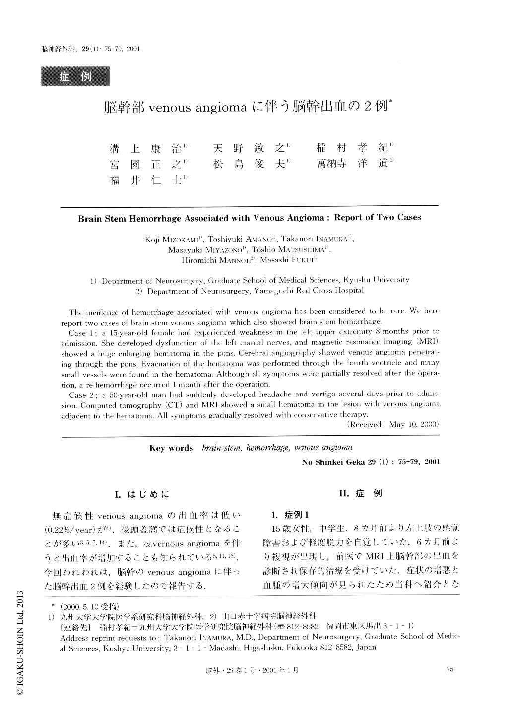
無症候性venous angiomaの出血率は低い(0.22%/year)が4),後頭蓋窩では症候性となることが多い3,5,7,14).また,cavernous angiomaを伴うと出血率が増加することも知られている5,11,16).今回われわれは,脳幹のvenous angiomaに伴った脳幹出血2例を経験したので報告する.
The incidence of hemorrhage associated with venous angioma has been considered to be rare. We here report two cases of brain stem venous angioma which also showed brain stem hemorrhage.
Case 1 ; a 15-year-old female had experienced weakness in the left upper extremity 8 months prior to admission. She developed dysfunction of the left cranial nerves, and magnetic resonance imaging (MRI) showed a huge enlarging hematoma in the pons. Cerebral angiography showed venous angioma penetrat- ing through the pons. Evacuation of the hematoma was performed through the fourth ventricle and many small vessels were found in the hematoma. Although all symptoms were partially resolved after the opera-tion, a re-hemorrhage occurred 1 month after the operation.
Case 2; a 50-year-old man had suddenly developed headache and vertigo several clays prior to admis-sion. Computed tomography (CT) and MRI showed a small hematoma in the lesion with venous angiomaadjacent to the hematoma. All symptoms gradually resolved with conservative therapy.

Copyright © 2001, Igaku-Shoin Ltd. All rights reserved.


