Japanese
English
- 有料閲覧
PPV購入案内
1,320円 (1,200円+税10%) こちらは、557ページから562ページの文献です。購入には医書.jp本会員登録が必要です。
会員登録完了後に改めて上記「購入サイトへ」を選択してください。
または「購入サイトへ」選択後に「購入ログイン」、「新規会員登録」の順に進んでください。
会員登録について詳しくはをご確認ください。
- Abstract 文献概要
- 1ページ目 Look Inside
I.はじめに
悪性線維性組織球腫(malignant fibrous histiocytoma以下MFHと略す)は,WHOの定義では一部,多形性を示す異型組織球と線維芽様細胞からなり,storiformpatternをとる悪性腫瘍とされる.軟部組織に発生する悪性新生物の10.5-21.6%を占めるとされ,主に四肢,後腹膜に好発する3,22).しかし,頭蓋内に原発した例は文献上散見するのみで,極めて稀である.今回,われわれは腫瘍出血により発症した頭蓋内原発と思われたMFHの1例を経験したので若干の文献的考察を加えて報告する.
Malignant fibrous histiocytoma (MFH) is common among the soft-tissue sarcoma and especially occurs in the retroperitoneum and the four extremities. However primary intracranial MFH is an extremely rare occur-rence.
A case with primary intracranial MFH associatedwith peritumoral hemorrhage is reported.
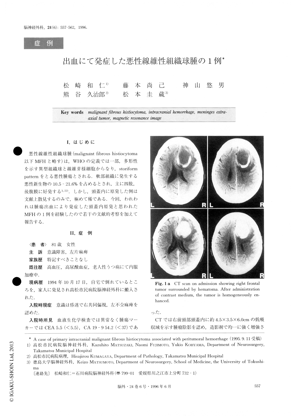
A 81-year-old woman was admitted to our depart-ment with impaired consciousness and left hemiparesis. On admission, she was drowsy. Computed tomography (CT) scans revealed a low density mass lesion of 4.5×3.5×6.0cm in diameter, which was surrounded by a high density area suggesting hematoma, in the right frontal lobe adjacent to the falx cerebri. The low densi-ty mass was homogeneously enhanced by contrast medium. Magnetic resonance imaging revealed that the mass was hypointense on T1 weighted image and hyperintense on T2 weighted image. After administra-tion of Gd-DTPA, the mass was homogeneously en-hanced and the falx cerebri was also enhanced at the site of attachment. Right carotid angiogram showed a tumor stain fed by the anterior cerebral artery. On op-eration, bleeding was encountered between the tumor and the falx cerebri on opening the dura mater. Hema-toma existed between the tumor and the normal cere-bral parenchyma. The tumor was sharply demarcated and didn't invade the normal cerebral parenchyma or the falx cerebri, so we considered that the tumor origin-ated from the meninges. Histologically, pleomorphic spindle cells in atypical storiform pattern were observed. Multinucleated giant cells were also seen. This case suggested to us that primary intracranial MFH may be associated with intracranial hemorrhage.
Copyright © 1996, Igaku-Shoin Ltd. All rights reserved.