Japanese
English
- 有料閲覧
PPV購入案内
1,320円 (1,200円+税10%) こちらは、183ページから188ページの文献です。購入には医書.jp本会員登録が必要です。
会員登録完了後に改めて上記「購入サイトへ」を選択してください。
または「購入サイトへ」選択後に「購入ログイン」、「新規会員登録」の順に進んでください。
会員登録について詳しくはをご確認ください。
- Abstract 文献概要
- 1ページ目 Look Inside
I.はじめに
1988年にDaumas-Duportにより提唱されたdys—embryoplastic neuroepithelial tumor(DNT)は,テント上の大脳皮質に発生するmixed neuroglial tumorの一型である.Daumas-Duportらの報告ではDNTとneuro—fibromatosis type 1(NF−1)などの母斑症との合併例はなかったと記載されており1,2),その後DNTの報告例が集積されているが,母斑症を合併したDNTは現在でも見あたらない1-8,11,12).しかしながら両者共に発生学的に胎生期の異常を基盤としており9,10),その合併症はDNTの発生起源を考える上で興味深い.最近われわれはNF−1を合併していたDNTの1例を経験したので報告する.
Dysembryoplastic neuroepithelial tumor (DNT) in a newly proposed mixed neuroglial tumor in the cerebral cortex. However, DNT associated with phacomatosis has mostly been considered exceptional. In this paper, a case of DNT associated with neurofibromatosis type 1 is reported.
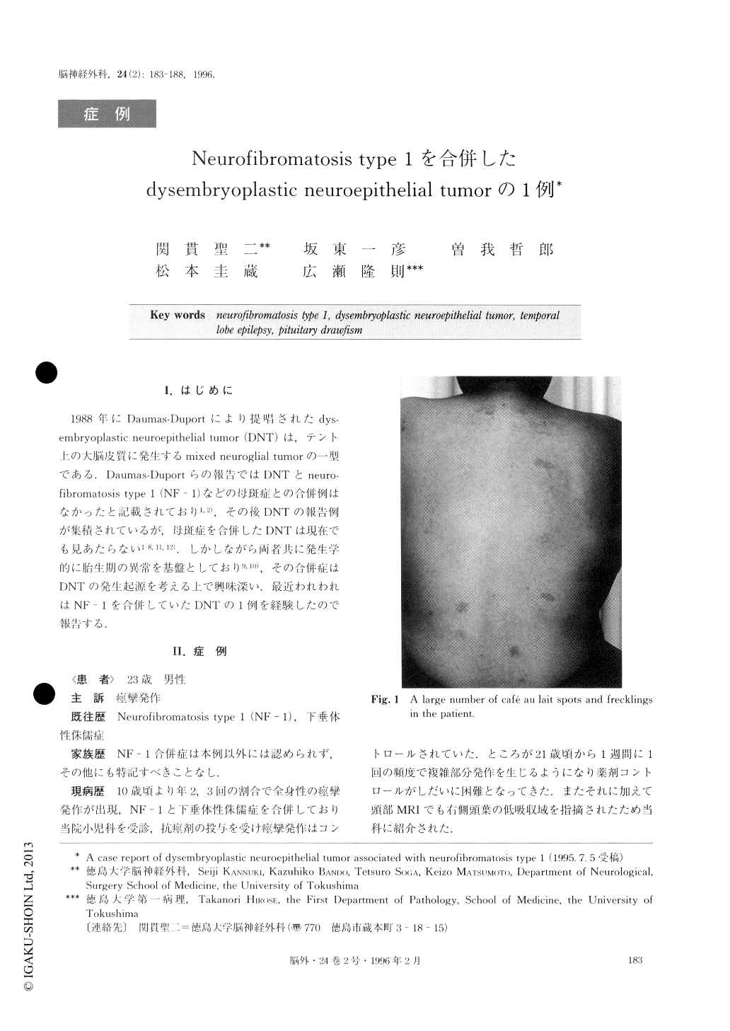
A 23-year-old male was admitted to our hospital complaining of intractable complex-partial seizure. He had a history of neurofibromatosis type 1 (NF-1) and pituitary dwarfism. On general physiological examina-tion, many cafe au lait spots and freckling could be noted, showing that the case was neurofibromatosis type 1. In addition, neurological examination showed no abnormal findings. MR images revealed a small area of abnormal intensity on the right temporal. This re-gion showed high intensity on T2 weighted image and low intensity without enhancement on T1 weighted im-age. On electroencephalography (EEG), an epileptic spike focus was demonstrated in the right temporal lobe. So, lobectomy was perfomed for control of epilepsy. Postoperative course was uneventful and without seizure. On histological examination, the tumor was composed of three different components: specific glioneural element, foci of oligodendrocyte-like cell, and cortical dysplasia. All of these findings were consistent with the definition of DNT by Daumas-Duport et al, except for the association with NF-1. Although re-ported cases of DNT associated with FN-1 are excep-tionally rare, both DNT and NF-1 originate from mal- development of the fetal central nervous system. It is very interesting that our case indicated the possibility of co-existence of both diseases.
Copyright © 1996, Igaku-Shoin Ltd. All rights reserved.