Japanese
English
- 有料閲覧
- Abstract 文献概要
- 1ページ目 Look Inside
I.はじめに
若年者のてんかん患者でCT上mass effectのないcysticなlow density areaを見た場合,porencephaly,arachnoid cyst, infarction後の変化などを考えるが,時にこの様な症例がsolidなlow grade gliomaであることがあり注意を要する5-13).CT上cysticなlow densityareaを示すテント上のlow grade gliomaの中で,最近Daumas-Duportらは,従来の分類にうまく当てはまらない特異な臨床病理学的特徴を有する一連の症例を報告し,これらをdysembryoplastic neuroepithelial tumorと名付けた4).われわれはこれに相当すると思われる1例を経験したので報告する.
Abstract
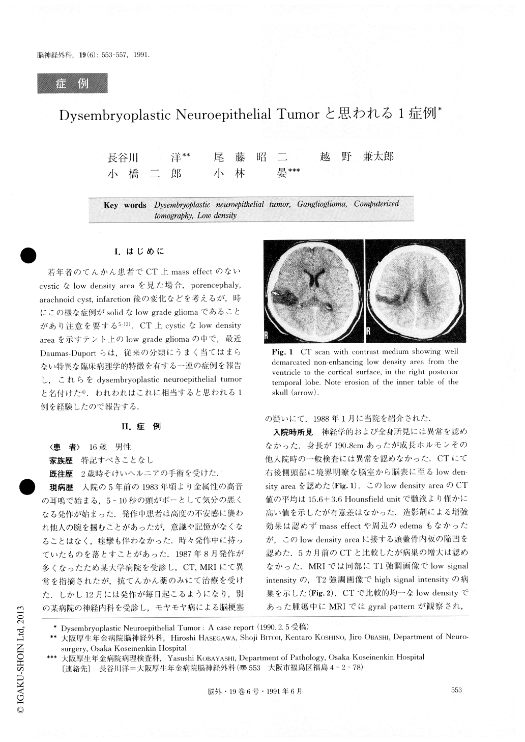
The authors report a case of dysembryoplastic neuroepithelial tumor which is a new entity of glial tumor proposed by Daumas-Duport et al. A 16-year-old male was admitted to our hospital with a 5-year history of uncontrollable complex partial seizure. CT scan showed a non-enhanced homogeneous low density area without mass effect, simulating old infarction or porencephalic cyst in the right posterior temporal lobe. The inner table of the skull over the lesion was eroded. The lesion showed low signal intensity in T1 weighted MR image and high signal intensity in T2 image. Cra-niotomy disclosed greyish soft solid tumor without cyst. Histologically, the tumor contained multiple cellular nodules in the microcystic astrocytic part which con-tained neurons. After the surgery the patient was free from the seizure.
Dysembryoplastic neuroepithelial tumor is found in young patients with intractable partial seizures. It is characterized by pseudocystic well-demarcated low den-sity appearance on CT scan. Histologically, it is an in-tracortical multinodular heterogeneous tumor which, is surgically treatable with favorable prognosis. For dif-ferential diagnosis, this tumor must be recognized in the list of low-density intracranial lesions found during CT scan.

Copyright © 1991, Igaku-Shoin Ltd. All rights reserved.


