Japanese
English
- 有料閲覧
PPV購入案内
1,320円 (1,200円+税10%) こちらは、1057ページから1061ページの文献です。購入には医書.jp本会員登録が必要です。
会員登録完了後に改めて上記「購入サイトへ」を選択してください。
または「購入サイトへ」選択後に「購入ログイン」、「新規会員登録」の順に進んでください。
会員登録について詳しくはをご確認ください。
- Abstract 文献概要
- 1ページ目 Look Inside
I.はじめに
小脳橋角部の脂肪腫は比較的稀であるが,われわれは最近,隣接動脈の奇形を伴った小脳橋角部脂肪腫の1例を経験した.本症例では脂肪腫内を走行する脳神経や血管がMRI上描出されるなど画像上も興味ある所見を得たので若干の文献的考察を加えて報告する.
A case was reported of cerebellopontine angle lipoma with vascular anomaly which was thought to he the remnant of fetal anastomosis of the posterior inferior cerebellar artery (PICA) and of the anterior inferior cerebellar artery (AICA).
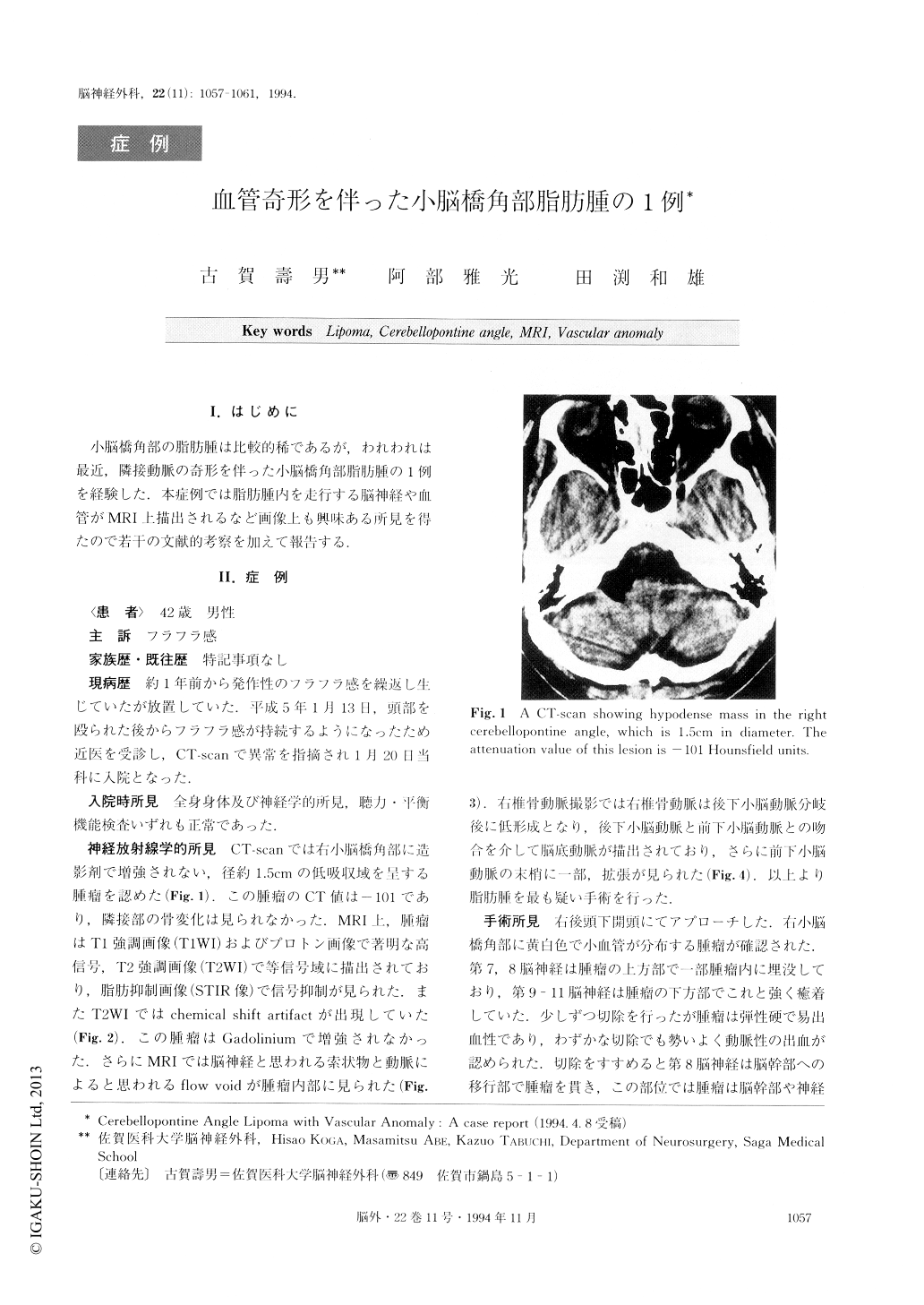
A 42 year-old man had been suffering from intermit-tent dizziness for a year. Worsening of dizziness after mild head trauma led him to visit a doctor who pointed out a cerebellopontine angle lesion. On admission to our hospital, neurological examination revealed no de-ficit. Computerized tomography (CT) scanning showed a low density mass in the right cerebellopontine angle, which was 1.5cm in diameter and showed no enhance-ment with intravenous contrast material. The attenua-tion value on CT of this lesion was -101 Hounsfield units. MRI showed a homogeneous mass, which was markedly hyperintense on T1 weighted image and pro-ton image, isointense with chemical shift artifact on T2 weighted image and markedly hypointense on short TIIR (STIR) image. Signal characteristics on MRI were consistent with those of lipoma. Interestingly, MRI de-monstrated the cranial nerves and vessels penetrating small lesion. The vertebral angiograms revealed the anastomosis of PICA and AICA with segmentally hypoplastic vertebral artery on the right side and the dilatation of the right distal AICA. Through a right suboccipital approach a yellowish mass in the right cerebellopontine angle was resected partially (about 50%). Only partial resection was possible because of the bleeding from small arteries and the tight adhesion to cranial nerves and brain stem. Histological findings of the surgical specimen were consistent with those of a lipoma. Postoperative neurological examination showed no neurological deficit. The patient has been completely free from dizziness since the surgery. Intracranial lipomas have been considered as a con-genital anomaly, which develops after 5 to 8 weeks of gestation, whereas anastomosis of the PICA and AICA normally vanishes after 6 to 8 weeks of gestation. As the number of reports regarding intracranial lipomas associated with vascular anomalies increases, we pre-sume there is a common etiology or a causal relation between these two factors.
Copyright © 1994, Igaku-Shoin Ltd. All rights reserved.