Japanese
English
- 有料閲覧
- Abstract 文献概要
- 1ページ目 Look Inside
- 参考文献 Reference
Ⅰ.はじめに
脊索腫は,胎生期の脊索組織の遺残から発生する腫瘍と考えられ7,11,13),約35%は頭蓋内,特に斜台先端部に発生する1,3,8,9,11,13).
われわれは,過去2回の手術で下垂体腺腫と診断したトルコ鞍内原発軟骨様脊索腫の1例を経験した.初期診断の誤りを再考し,かつトルコ鞍内原発脊索腫について考察を加え報告する.
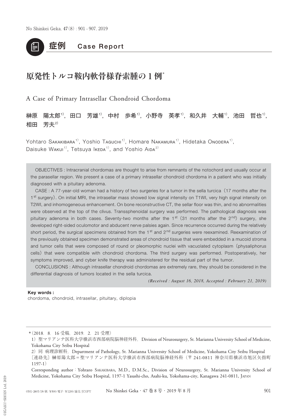
OBJECTIVES:Intracranial chordomas are thought to arise from remnants of the notochord and usually occur at the parasellar region. We present a case of a primary intrasellar chondroid chordoma in a patient who was initially diagnosed with a pituitary adenoma.
CASE:A 77-year-old woman had a history of two surgeries for a tumor in the sella turcica(17 months after the 1st surgery). On initial MRI, the intrasellar mass showed low signal intensity on T1WI, very high signal intensity on T2WI, and inhomogeneous enhancement. On bone reconstructive CT, the sellar floor was thin, and no abnormalities were observed at the top of the clivus. Transsphenoidal surgery was performed. The pathological diagnosis was pituitary adenoma in both cases. Seventy-two months after the 1st(31 months after the 2nd)surgery, she developed right-sided oculomotor and abducent nerve palsies again. Since recurrence occurred during the relatively short period, the surgical specimens obtained from the 1st and 2nd surgeries were reexamined. Reexamination of the previously obtained specimen demonstrated areas of chondroid tissue that were embedded in a mucoid stroma and tumor cells that were composed of round or pleomorphic nuclei with vacuolated cytoplasm(physaliphorus cells)that were compatible with chondroid chordoma. The third surgery was performed. Postoperatively, her symptoms improved, and cyber knife therapy was administered for the residual part of the tumor.
CONCLUSIONS:Although intrasellar chondroid chordomas are extremely rare, they should be considered in the differential diagnosis of tumors located in the sella turcica.

Copyright © 2019, Igaku-Shoin Ltd. All rights reserved.


