Japanese
English
- 有料閲覧
PPV購入案内
1,320円 (1,200円+税10%) こちらは、735ページから738ページの文献です。購入には医書.jp本会員登録が必要です。
会員登録完了後に改めて上記「購入サイトへ」を選択してください。
または「購入サイトへ」選択後に「購入ログイン」、「新規会員登録」の順に進んでください。
会員登録について詳しくはをご確認ください。
- Abstract 文献概要
- 1ページ目 Look Inside
I.はじめに
頭部の腫瘤性病変は,日常診療においてしばしば遭遇され,外来にて治療されていることが多い.しかし,腫瘤の大きさや性状によっては鑑別診断が必要な症例も存在する.
今回,われわれは長年の経過のうちに増大してきた,成人の後頭部皮下に巨大腫瘤を形成した類上皮腫の1例を経験したので,鑑別診断を含めて文献的考察を加えて報告する.
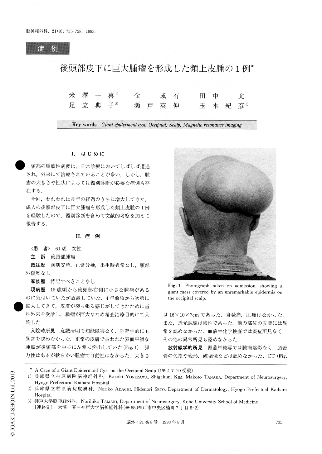
The authors report a case of a 61-year-old woman presenting with a giant mass on the occipital scalp. The patient had no neurological deficits and was of normal in-telligence. The lesion was soft and covered by an unre-markable epidermis. The preoperative radiologic evalua-tion was made by CT, MR Imaging, and by cerebral angi ogram. The mass produced an inhomogeneously low-intensity signal involving also a high-intensity signal. It was shown, on the T1 weighted image, that it did not communicate with the intracranial space, and there was no gadolinium-enhanced lesion. MR Imaging was supe-rior to CT in the evaluation of the giant mass on the scalp and was particularly useful in surgical planning. At sur-gical resection, a soft fluffy, white-colored tumor which measured 16×10×7cm was totally removed. Patholo-gical diagnosis of the tumor was epidermoid cyst.
Copyright © 1993, Igaku-Shoin Ltd. All rights reserved.