Japanese
English
- 有料閲覧
PPV購入案内
1,320円 (1,200円+税10%) こちらは、157ページから159ページの文献です。購入には医書.jp本会員登録が必要です。
会員登録完了後に改めて上記「購入サイトへ」を選択してください。
または「購入サイトへ」選択後に「購入ログイン」、「新規会員登録」の順に進んでください。
会員登録について詳しくはをご確認ください。
- Abstract 文献概要
- 1ページ目 Look Inside
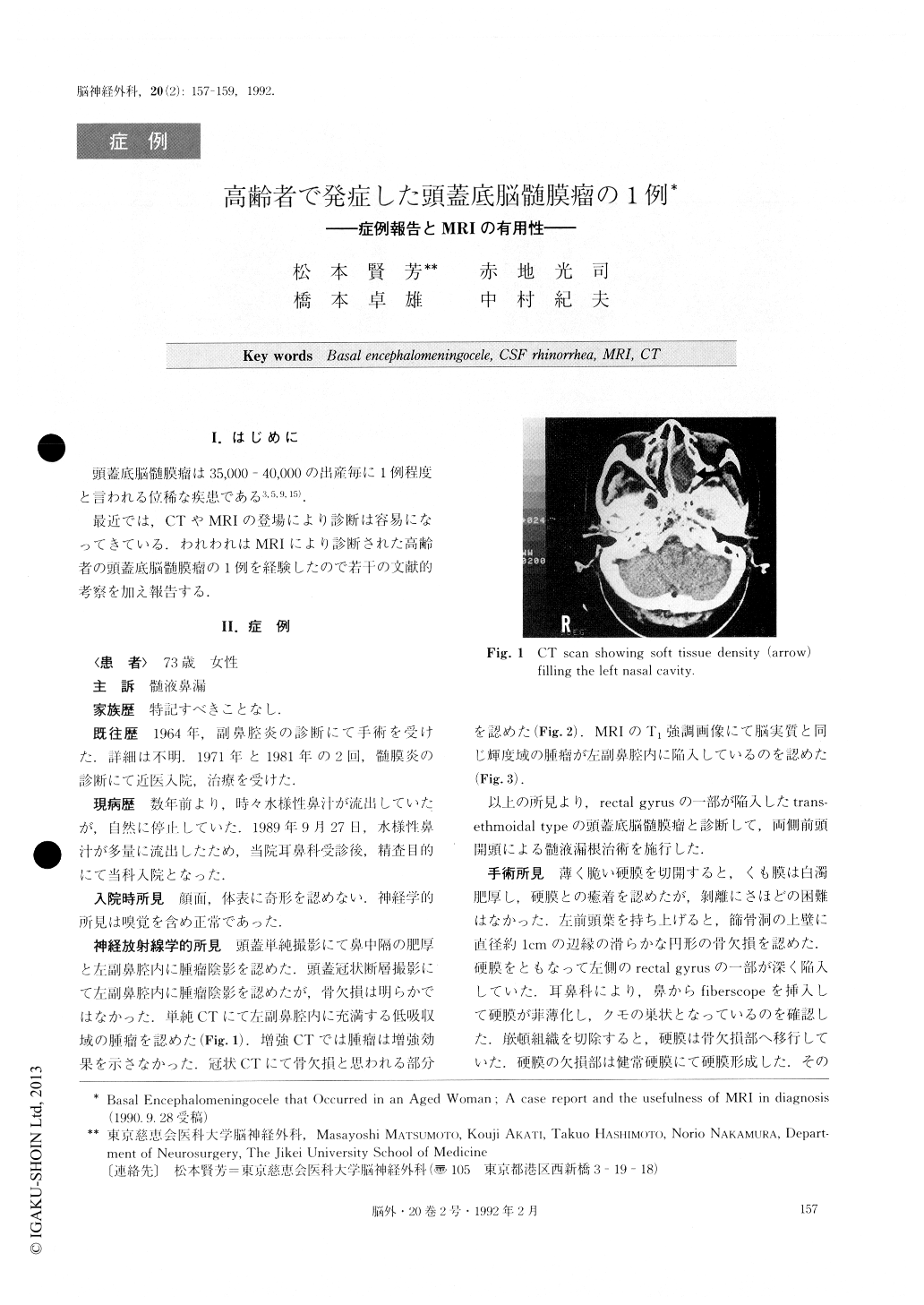
I.はじめに
頭蓋底脳髄膜瘤は35,000-40,000の出産毎に1例程度と言われる位稀な疾患である3,5,9,15).
最近では,CTやMRIの登場により診断は容易になってきている.われわれはMRIにより診断された高齢者の頭蓋底脳髄膜瘤の1例を経験したので若干の文献的考察を加え報告する.
The usefulness of MRI in diagnosis of a basal en-cephalomeningocele case is reported. A 72-year-old woman with a several-year history of occasional watery rhinorrhea and previous histories of meningitis at the ages of 55 and 65 is the subject of this report initially. After referring to the department of otorhinolaryngolo-gy, she was admitted to our department for close ex-amination.
CT scan showed only a mass forming a cyst in the nasal cavity, but MRI demonstrated part of the left rec-tal gyrus invaginating into the ethmoid sinus and nasal cavity.
However, from the next day the patient developed left hemi-paresis. Right carotid angiogram on the 17th day after the injury revealed multiple segmental arterial narrow-ing in the right anterior cerebral artery (ACA) and middle cerebral artery (MCA). We diagnosed a post-traumatic delayed cerebral arterial spasm. CT scan re-vealed low density areas in the right ACA and MCA territory.
The pathogenesis of posttraumatic delayed arterial spasm is not yet well known. Now, four theories have been suggested as follows:① Subarachnoid hemor-rhage,②Direct mechanical injury to the arterial wall,③ Hypothalamus dysfunction, and④Disturbed auto-regulation
In our case, three important factors are suggested. The first is direct injury to the artery, the second is cerebral contusion, and the third is subdural effusion.
Copyright © 1992, Igaku-Shoin Ltd. All rights reserved.