Japanese
English
- 有料閲覧
- Abstract 文献概要
- 1ページ目 Look Inside
I.はじめに
頭部外傷後の脳血管攣縮の発生頻度は5-47.7%とされ11),報告によって大きな差がある.しかし,外傷性脳血管攣縮で臨床症状を呈することは比較的少なく,そのメカニズムの究明もまだ十分なされていない.また,症候性脳血管攣縮は予後に影響する因子であることから,その発生を予期し,早期に対処することが必要とおもわれる.今回,われわれは頭部外傷後に前大脳動脈と中大脳動脈に脳血管攣縮を来した症例を経験したので若十の文献的考察を加えて報告する.
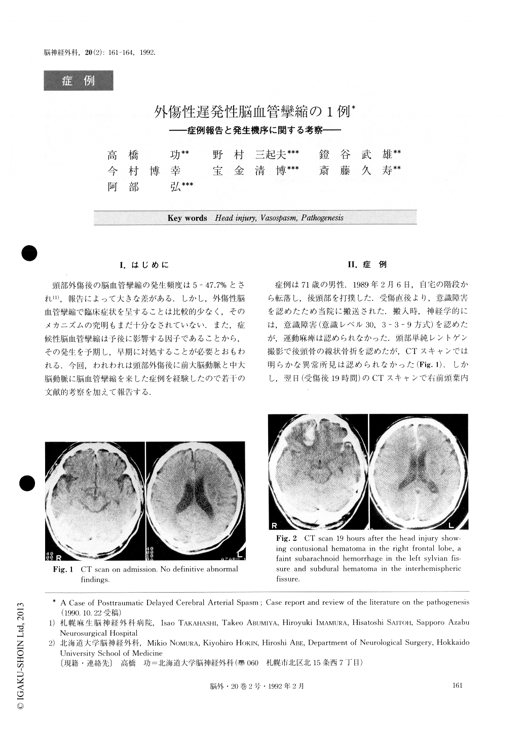
A case of posttraumatic delayed cerebral arterial spasm is presented. A 71-year-old man was admitted to our hospital with head injury. Neurological examination on admission only revealed consciousness disturbance (Japan Coma Scale 30). CT scan 19 hours after the in-jury demonstrated a contusional hematoma in the right frontal lobe, faint subarachnoid hemorrhage in the left sylvian fissure and subdural hematoma in the interhe-mispheric fissure. His consciousness was disturbed on the 14th day. CT scan demonstrated a left subdural effusion, which was surgically evacuated. However, from the next day the patient developed left hemi-paresis. Right carotid angiogram on the 17th day after the injury revealed multiple segmental arterial narrow-ing in the right anterior cerebral artery (ACA) and middle cerebral artery (MCA) . We diagnosed a post-traumatic delayed cerebral arterial spasm. CT scan re-vealed low density areas in the right ACA and MCA territory.
The pathogenesis of posttraumatic delayed arterial spasm is not yet well known. Now, four theories have been suggested as follows :①Subarachnoid hemor- rhage, ②Direct mechanical injury to the arterial wall, ③Hypothalamus dysfunction, and ④Disturbed auto-regulation
In our case, three important factors are suggested. The first is direct injury to the artery, the second is cerebral contusion, and the third is subdural effusion.

Copyright © 1992, Igaku-Shoin Ltd. All rights reserved.


