Japanese
English
- 有料閲覧
PPV購入案内
1,320円 (1,200円+税10%) こちらは、1161ページから1166ページの文献です。購入には医書.jp本会員登録が必要です。
会員登録完了後に改めて上記「購入サイトへ」を選択してください。
または「購入サイトへ」選択後に「購入ログイン」、「新規会員登録」の順に進んでください。
会員登録について詳しくはをご確認ください。
- Abstract 文献概要
- 1ページ目 Look Inside
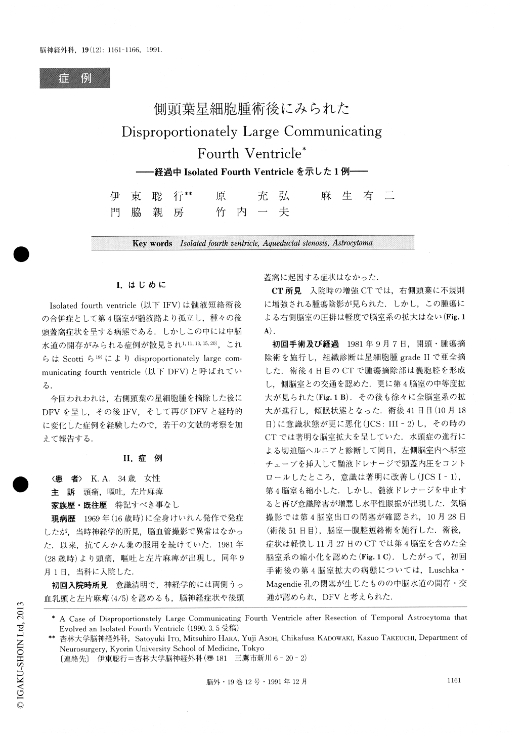
I.はじめに
Isolated fourth ventricle(以下IFV)は髄液短絡術後の合併症として第4脳室が髄液路より孤立し,種々の後頭蓋窩症状を呈する病態である.しかしこの中には中脳水道の開存がみられる症例が散見され1,11,13,15,20),これらはScottiら19)によりdisproportlonately large com—municating fourth ventricle(以下DFV)と呼ばれている.
今回われわれは,右側頭葉の星細胞腫を摘除した後にDFVを呈し,その後IFV,そして再びDFVと経時的に変化した症例を経験したので,若干の文献的考察を加えて報告する.
The isolation and enlargement of the fourth ventricle after a ventriculoperitoneal (V-P) shunt was classified as “isolated fourth ventricle (IFV) ”. The term, “dis-proportionately large communicating fourth ventricle (DFV) ” was first introduced by Scotti et al as being an enlarged fourth ventricle communicating with the third ventricle.
The authors present a case of DFV after the resec-tion of an astrocytoma. Upon recurrence of the tumor a second resection was carried out 5 years later. It was found that IFV had evolved because a cyst in the right temporal lobe was obstructing the aqueduct. After shunting of the tumor cyst, the aqueduct was again found to be patent and the fourth ventricle gradually decreased in size.
A 34-year-old female presented headache, nausea, and a mild left hemiparesis. An initial CT scan demon-strated a fourth ventricle of approximately normal size and a right temporal mass. The first craniotomy re-vealed an astrocytoma. A CT scan after the surgical procedure showed enlargement of all ventricles, espe-cially the fourth, resulting from the blockage of the foramina of Luschka and Magendie. The insertion of a V-P shunt was followed by a reduction in size of all ventricles. The diagnosis of DFV was thus confirmed because the fourth ventricle had a demonstrated com-munication with the third ventricle. After a second cra-niotomy for tumor recurrence five years later, a CT scan revealed the enlargement of the fourth ventricle and a cyst in the right temporal lobe. A metrizamide CT scan revealed that the cyst was isolated and an RI ventriculogram confirmed obstruction of the aqueduct. We diagnosed IFV and a cyst-peritoneal shunt was then carried out. After the shunt procedure, a bilateral sixth nerve palsy and limitation of upward gaze werenoted. A CT scan showed that the size of the cyst had decreased, while the fourth ventricle had not decreased in size. A repeat ventriculogram indicated a patent aqueduct, clearly demonstrating DFV. One year later, abnormal extra-ocular movements were improved, and recent CT scans and magnetic resonance images show a normal sized fourth ventricle.
Copyright © 1991, Igaku-Shoin Ltd. All rights reserved.