Japanese
English
- 有料閲覧
PPV購入案内
1,320円 (1,200円+税10%) こちらは、897ページから902ページの文献です。購入には医書.jp本会員登録が必要です。
会員登録完了後に改めて上記「購入サイトへ」を選択してください。
または「購入サイトへ」選択後に「購入ログイン」、「新規会員登録」の順に進んでください。
会員登録について詳しくはをご確認ください。
- Abstract 文献概要
- 1ページ目 Look Inside
I.はじめに
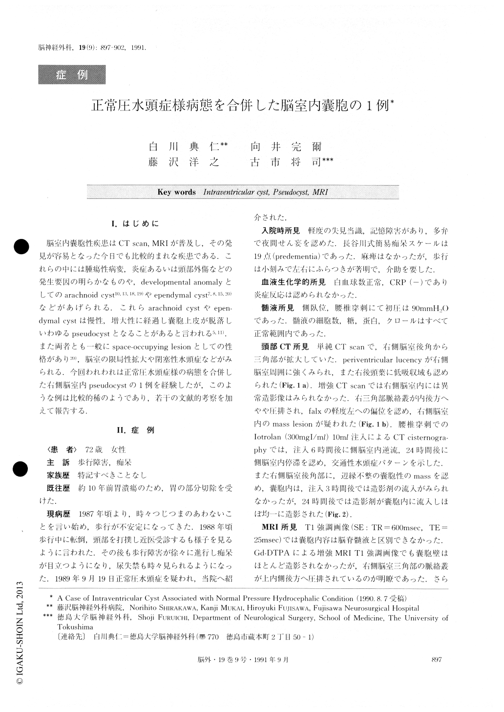
脳室内嚢胞性疾患はCT scan, MRIが普及し,その発見が容易となった今日でも比較的まれな疾患である.これらの中には腫瘍性病変,炎症あるいは頭部外傷などの発生要因の明らかなものや,developmental anomalyとしてのarachnoid cyst10,13,18,19)やependymal cyst7,8,15,20)などがあげられる.これらarachnoid cystやepen—dymai cystは慢性,増大性に経過し嚢胞上皮が脱落しいわゆるpseudocystとなることがあると言われる5,11).また両者とも一般にspace-occupying lesionとしての性格があり20),脳室の限局性拡大や閉塞性水頭症などがみられる.今回われわれは正常圧水頭症様の病態を合併した右側脳室内pseudocystの1例を経験したが,このような例は比較的稀のようであり,若干の文献的考察を加えて報告する.
Abstract
A case of intraventricular cyst associated with normal pressure hydrocephalic condition (NPHC) is reported.
A 72 year-old female, with 2-year-history of slowly progressing dementia and gait disturbance, was admitted to our hospital on September 19, 1989. On admission, she had mild dementia, unsteadiness of gait, andat times urinary incontinence. Cerebrospinal fluid (CSF) pressure was found to be 90mmH2O by lumbar tap. Plain computed tomographic (CT) scan and T1-weighted magnetic resonance image (MRI) showed asymmetrical enlargement of the trigon and posterior horn of the right lateral ventricle. CT cisternography showed a cyst in the trigon and in the posterior horn of the right lateral ventricle. T1-weighted MRI with Gd-DTPA demonstrated no enhancement of the cyst wall, and there was superior and posteromedial displacement of the choroid plexus at the trigon of the right lateral ventricle.
The patient was diagnosed as having an intraventricular cyst in the right trigon with NPHC. Ventriculo-peritoneal shunt and partial removal of the cyst were performed. Histological examination of the cyst wall revealed collagenous strands and no epithelial cells.
Developmental intracranial cysts, especially arachnoid or ependymal cysts, occasionally lack an epithelial layer, so their histological diagnosis is difficult. This case was considered to be an arachnoid cyst because there was adhesion between the cyst and the choroid plexus in the right trigon, and superior, posteromedial displacement of the choroid plexus, which indicated extension of the cyst from the extracerebral to the intracerehral region. NPHC was considered to be due to disturbance of CSF. circulation caused by gradual expansion of the cyst.
Copyright © 1991, Igaku-Shoin Ltd. All rights reserved.