Japanese
English
- 有料閲覧
- Abstract 文献概要
- 1ページ目 Look Inside
I.はじめに
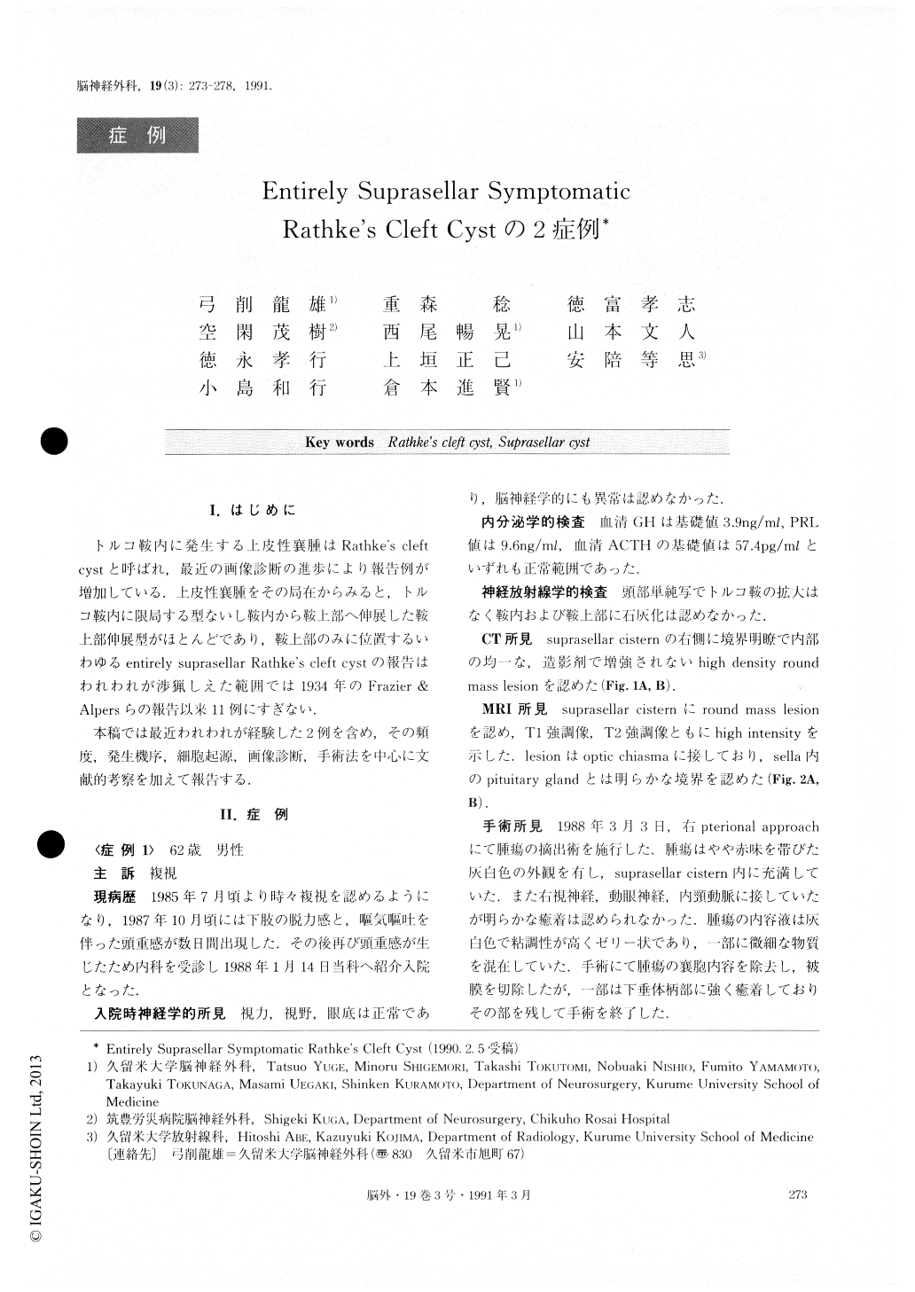
トルコ鞍内に発生する上皮性襄腫はRathke's cleftcystと呼ばれ,最近の画像診断の進歩により報告例が増加している.上皮性襄腫をその局在からみると,トルコ鞍内に限局する型ないし鞍内から鞍上部へ伸展した鞍上部仲展型がほとんどであり,鞍上部のみに位置するいわゆるentirely suprasellar Rathke's cleft cystの報告はわれわれが渉猟しえた範囲では1934年のFrazier & Alpersらの報告以来11例にすぎない.
本稿では最近われわれが経験した2例を含め,その頻度,発生機序,細胞起源,画像診断,手術法を中心に文献的考察を加えて報告する.
Abstract
Two rare cases of entirely suprasellar Rathke's cleft cyst were reported.
Case 1. A 62-year-old man was admitted to our hos-pital on the 14th of January, 1988, complaining of headache and diplopia.
A plain skull x-ray showed the sella turicica was nor-mal. CT scan and MRI demonstrated a lesion mass lo-cated entirely in the suprasellar cistern. Right fronto-temporal craniotomy was performed, and the cyst wall was resected subtotally. Microscopic sections of cyst wall showed ciliated single layer with focal stratified epithelium.
Case 2. A 51-year-old man was hospitalised complain-ing of visual impairment in the left eye. Endocrinologi-cal examination showed no abnormalities. CT and MRI demnostrated a lesion mass located entirely in the su-prasellar region. Right frontotemporal craniotomy was performed. The mass was opened and a large amount of yellowish fluid was released.
Histologically, the specimens were simple ciliated cuboidal epihelium. Postoperative courses of these pa-tients were uneventful. The findings on CT and MRI of the cases located entirely in the suprasellar region were varied. The histopathogenesis and embryological pathogenesis of Rathke's cleft cyst in the literature, particularly the entirely suprasellar type, were dis-cussed.

Copyright © 1991, Igaku-Shoin Ltd. All rights reserved.


