Japanese
English
- 有料閲覧
PPV購入案内
1,320円 (1,200円+税10%) こちらは、43ページから57ページの文献です。購入には医書.jp本会員登録が必要です。
会員登録完了後に改めて上記「購入サイトへ」を選択してください。
または「購入サイトへ」選択後に「購入ログイン」、「新規会員登録」の順に進んでください。
会員登録について詳しくはをご確認ください。
- Abstract 文献概要
- 1ページ目 Look Inside
- 参考文献 Reference
Point
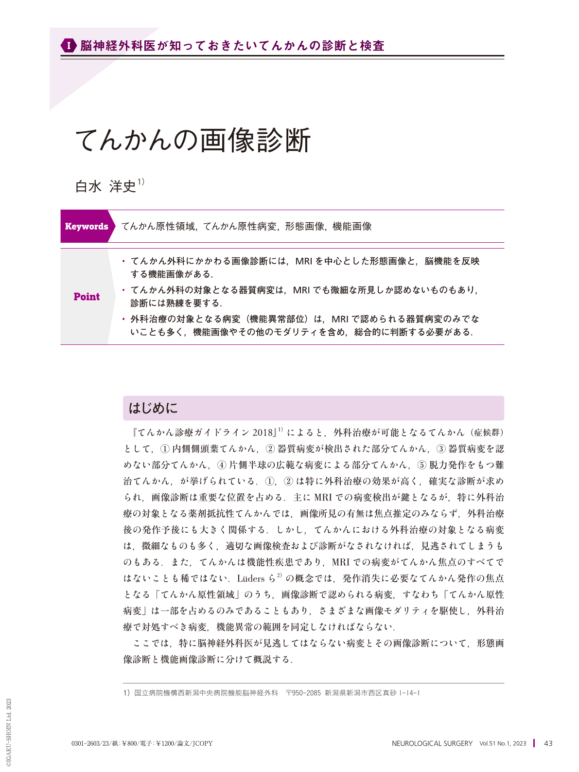
・てんかん外科にかかわる画像診断には,MRIを中心とした形態画像と,脳機能を反映する機能画像がある.
・てんかん外科の対象となる器質病変は,MRIでも微細な所見しか認めないものもあり,診断には熟練を要する.
・外科治療の対象となる病変(機能異常部位)は,MRIで認められる器質病変のみでないことも多く,機能画像やその他のモダリティを含め,総合的に判断する必要がある.
Neuroimaging is commonly used for presurgical evaluation in epilepsy surgery. Neuroimaging for epilepsy includes structural and functional neuroimaging. Lesions detected by structural neuroimaging are crucial to determine the indication of epilepsy surgery, as well as to predict seizure outcomes, as patients with MRI-visible lesions are likely to have better seizure outcomes. However, MRI lesions sometimes show very faint findings; therefore, the diagnosis of structural neuroimaging requires sophisticated skills. Moreover, the epilepsy focus should not only involve the MRI-visible lesion, but also the surrounding tissue with abnormal neuronal function. The MRI-lesion, which is almost the same as that epileptogenic lesion, is a part of the epileptogenic zone. Surgical strategy should be conducted by comprehensive evaluation including neuroimaging in addition to other modalities.
Copyright © 2023, Igaku-Shoin Ltd. All rights reserved.