Japanese
English
- 有料閲覧
PPV購入案内
1,320円 (1,200円+税10%) こちらは、724ページから736ページの文献です。購入には医書.jp本会員登録が必要です。
会員登録完了後に改めて上記「購入サイトへ」を選択してください。
または「購入サイトへ」選択後に「購入ログイン」、「新規会員登録」の順に進んでください。
会員登録について詳しくはをご確認ください。
- Abstract 文献概要
- 1ページ目 Look Inside
- 参考文献 Reference
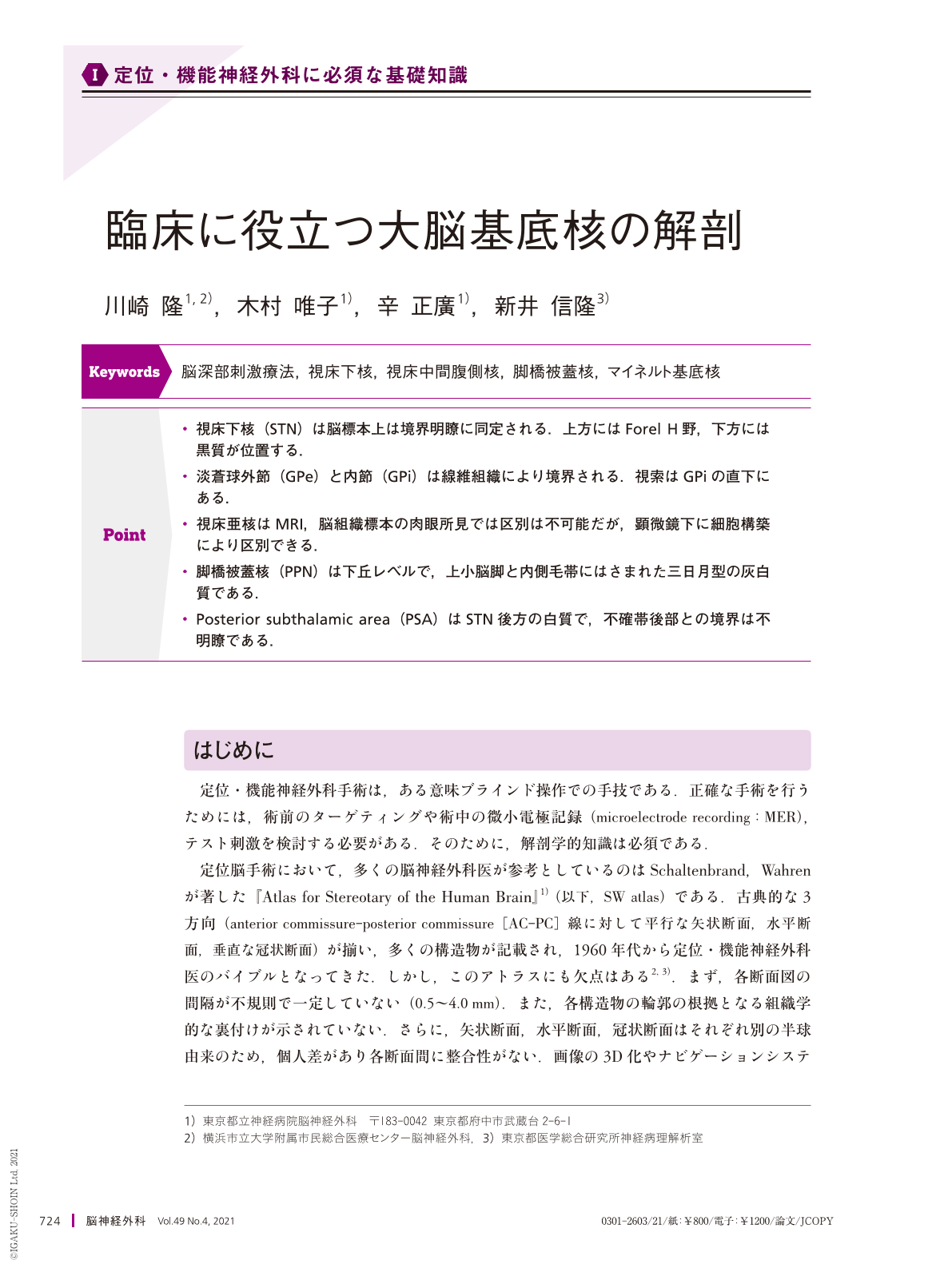
Point
・視床下核(STN)は脳標本上は境界明瞭に同定される.上方にはForel H野,下方には黒質が位置する.
・淡蒼球外節(GPe)と内節(GPi)は線維組織により境界される.視索はGPiの直下にある.
・視床亜核はMRI,脳組織標本の肉眼所見では区別は不可能だが,顕微鏡下に細胞構築により区別できる.
・脚橋被蓋核(PPN)は下丘レベルで,上小脳脚と内側毛帯にはさまれた三日月型の灰白質である.
・Posterior subthalamic area(PSA)はSTN後方の白質で,不確帯後部との境界は不明瞭である.
Anatomical knowledge of target structures is essential in stereotactic functional neurosurgery. Thus, we created a three-dimensional(3D)atlas comprising frozen sections and histologically stained slides prepared from cadaveric brains. Herein, we describe the anatomical information of stereotactic functional neurosurgery targets gained from our atlas.
The subthalamic nucleus(STN)was found to be clearly enclosed by neural fibers with high neuronal density. Based on our 3D models, the mean penetration length of deep brain stimulation leading into the STN was 6.6 mm.
The globus pallidus was found to be clearly divided into the grobus pallidus externus(GPe)and internus(GPi)by its neural fibers, and the optic tract was located below the GPi.
Although the thalamic lateral nuclear group(ventrooralis, ventrontermedius, and ventrocaudalis)could not be identified from either macroscopic frozen sections or MR images, these structures were clearly discernible from each other based on cell architecture(cell size and cell density)when viewed under a microscope. In contrast, distinguishing ventral and dorsal nuclei in humans is difficult.
In addition to the main targets of the basal ganglia, we also investigated the anatomy of other targets in detail(posterior subthalamic area, pedunclopontine nucleus, nucleus accumbens, and nucleus basalis of Meynert).
Overall, this anatomical knowledge from the atlas helps functional neurosurgeons interpret intraoperative microelectrode recording and MRI more precisely, helping facilitate more accurate surgeries.
Copyright © 2021, Igaku-Shoin Ltd. All rights reserved.