Japanese
English
- 有料閲覧
PPV購入案内
1,320円 (1,200円+税10%) こちらは、275ページから279ページの文献です。購入には医書.jp本会員登録が必要です。
会員登録完了後に改めて上記「購入サイトへ」を選択してください。
または「購入サイトへ」選択後に「購入ログイン」、「新規会員登録」の順に進んでください。
会員登録について詳しくはをご確認ください。
- Abstract 文献概要
- 1ページ目 Look Inside
I.はじめに
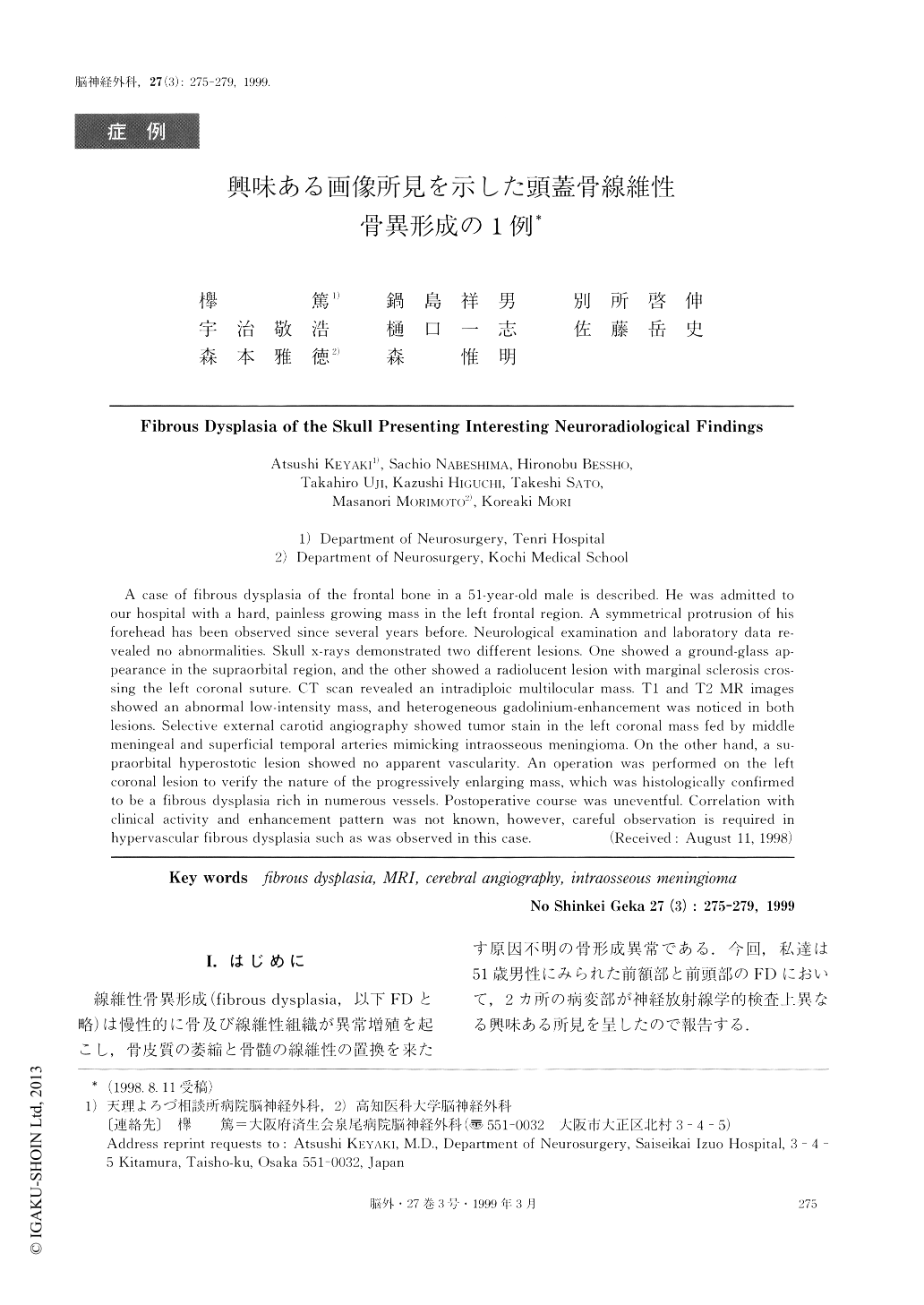
線維性骨異形成(fibrous dysplasia,以下FDと略)は慢性的に骨及び線維性組織が異常増殖を起こし,骨皮質の萎縮と骨髄の線維性の置換を来たす原因不明の骨形成異常である.今回,私達は51歳男性にみられた前額部と前頭部のFDにおいて,2ヵ所の病変部が神経放射線学的検査上異なる興味ある所見を呈したので報告する.
A case of fibrous dysplasia of the frontal bone in a 51-year-old male is described. He was admitted toour hospital with a hard, painless growing mass in the left frontal region. A symmetrical protrusion of hisforehead has been observed since several years before. Neurological examination and laboratory data re-vealed no abnormalities. Skull x-rays demonstrated two different lesions. One showed a ground-glass ap-pearance in the supraorbital region, and the other showed a radiolucent lesion with marginal sclerosis cros-sing the left coronal suture. CT scan revealed an intradiploic multilocular mass. T1 and T2 MR imagesshowed an abnormal low-intensity mass, and heterogeneous gadolinium-enhancement was noticed in bothlesions. Selective external carotid angiography showed tumor stain in the left coronal mass fed by middlemeningeal and superficial temporal arteries mimicking intraosseous meningioma. On the other hand, a su-praorbital hyperostotic lesion showed no apparent vascularity. An operation was performed on the leftcoronal lesion to verify the nature of the progressively enlarging mass, which was histologically confirmedto be a fibrous dysplasia rich in numerous vessels. Postoperative course was uneventful. Correlation withclinical activity and enhancement pattern was not known, however, careful observation is required inhypervascular fibrous dysplasia such as was observed in this case.
Copyright © 1999, Igaku-Shoin Ltd. All rights reserved.