Japanese
English
- 有料閲覧
PPV購入案内
1,320円 (1,200円+税10%) こちらは、771ページから781ページの文献です。購入には医書.jp本会員登録が必要です。
会員登録完了後に改めて上記「購入サイトへ」を選択してください。
または「購入サイトへ」選択後に「購入ログイン」、「新規会員登録」の順に進んでください。
会員登録について詳しくはをご確認ください。
- Abstract 文献概要
- 1ページ目 Look Inside
- 参考文献 Reference
Ⅰ.はじめに
側方経路腰椎椎体間固定術(lateral lumbar interbody fusion:LLIF)は,後腹膜経路に椎体側方より腰椎椎体間固定を行う手技であるが,出血量の減少,間接除圧効果,高い変形矯正力により手術侵襲が低減され,さまざまな病態に応用されるようになってきた15).特に後方椎体間固定術(posterior lumbar interbody fusion:PLIF)では,大量出血を伴うことがあるため避けられがちであった多椎間固定への道が広がった.一方,成人脊柱変形(adult spinal deformity:ASD)では,骨盤を含む脊柱矢状面アライメントの悪化が健康関連QOLを低下させると報告されているが2,11),近年急速に進む高齢化に伴いASD患者は増加し,その治療ニーズも高まりつつある.
ASD手術の目的は,良好なglobal balanceを獲得することであるが,適切な矯正と固定を得るために広範囲固定,骨切りなど高侵襲手術が必要であり,従前のオープン法による手術では,術中大量出血,術後創部感染などさまざまな周術期合併症が危惧される15).近年,最小侵襲脊椎安定術(minimally invasive spine stabilization:MISt)の普及や技術の発展に伴い,さまざまな疾患に応用されるようになってきた.LLIFと経皮的椎弓根スクリュー(percutaneous pedicle screw:PPS)を組み合わせたcircumferential minimally invasive surgery(cMIS)をASD手術に行い,出血量減少,輸血率減少,重篤な合併症が少ない,早期離床が可能などの利点が報告されている5).その反面,cMISは冠状面アライメントの改善には優れるが,矢状面アライメントの改善に乏しく,適応は症状がmildからmoderateな症例に限られ,重篤な矢状面アライメント不良例には適さないと指摘されてきた5).
今回われわれは,LLIFの1つであるoblique lumbar interbody fusion(OLIF)とPPSを用いたcMISでASDに対する矯正手術を行い,良好な結果を得たので,その詳細を報告する.
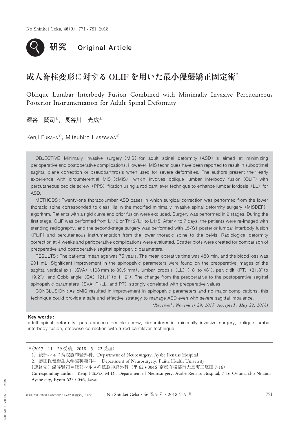
OBJECTIVE:Minimally invasive surgery(MIS)for adult spinal deformity(ASD)is aimed at minimizing perioperative and postoperative complications. However, MIS techniques have been reported to result in suboptimal sagittal plane correction or pseudoarthrosis when used for severe deformities. The authors present their early experience with circumferential MIS(cMIS), which involves oblique lumbar interbody fusion(OLIF)with percutaneous pedicle screw(PPS)fixation using a rod cantilever technique to enhance lumbar lordosis(LL)for ASD.
METHODS:Twenty-one thoracolumbar ASD cases in which surgical correction was performed from the lower thoracic spine corresponded to class IIIa in the modified minimally invasive spinal deformity surgery(MISDEF)algorithm. Patients with a rigid curve and prior fusion were excluded. Surgery was performed in 2 stages. During the first stage, OLIF was performed from L1/2 or Th12/L1 to L4/5. After 4 to 7 days, the patients were re-imaged with standing radiography, and the second-stage surgery was performed with L5/S1 posterior lumbar interbody fusion(PLIF)and percutaneous instrumentation from the lower thoracic spine to the pelvis. Radiological deformity correction at 4 weeks and perioperative complications were evaluated. Scatter plots were created for comparison of preoperative and postoperative sagittal spinopelvic parameters.
RESULTS:The patients' mean age was 75 years. The mean operative time was 488 min, and the blood loss was 901 mL. Significant improvement in the spinopelvic parameters were found on the preoperative images of the sagittal vertical axis(SVA)(108mm to 33.5 mm), lumbar lordosis(LL)(18°to 48°), pelvic tilt(PT)(31.8°to 19.2°), and Cobb angle(CA)(21.1°to 11.9°). The change from the preoperative to the postoperative sagittal spinopelvic parameters(SVA, PI-LL, and PT)strongly correlated with preoperative values.
CONCLUSION:As cMIS resulted in improvement in spinopelvic parameters and no major complications, this technique could provide a safe and effective strategy to manage ASD even with severe sagittal imbalance.
Copyright © 2018, Igaku-Shoin Ltd. All rights reserved.