Japanese
English
- 有料閲覧
PPV購入案内
1,320円 (1,200円+税10%) こちらは、811ページから817ページの文献です。購入には医書.jp本会員登録が必要です。
会員登録完了後に改めて上記「購入サイトへ」を選択してください。
または「購入サイトへ」選択後に「購入ログイン」、「新規会員登録」の順に進んでください。
会員登録について詳しくはをご確認ください。
- Abstract 文献概要
- 1ページ目 Look Inside
- 参考文献 Reference
Ⅰ.はじめに
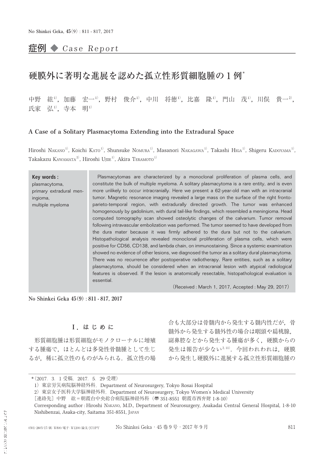
形質細胞腫は形質細胞がモノクローナルに増殖する腫瘍で,ほとんどは多発性骨髄腫として生じるが,稀に孤立性のものがみられる.孤立性の場合も大部分は骨髄内から発生する髄内性だが,骨髄外から発生する髄外性の場合は咽頭や扁桃腺,副鼻腔などから発生する腫瘍が多く,硬膜からの発生は報告が少ない2,11).今回われわれは,硬膜から発生し硬膜外に進展する孤立性形質細胞腫の1例を経験したので報告する.
Plasmacytomas are characterized by a monoclonal proliferation of plasma cells, and constitute the bulk of multiple myeloma. A solitary plasmacytoma is a rare entity, and is even more unlikely to occur intracranially. Here we present a 62-year-old man with an intracranial tumor. Magnetic resonance imaging revealed a large mass on the surface of the right fronto-parieto-temporal region, with extradurally directed growth. The tumor was enhanced homogenously by gadolinium, with dural tail-like findings, which resembled a meningioma. Head computed tomography scan showed osteolytic changes of the calvarium. Tumor removal following intravascular embolization was performed. The tumor seemed to have developed from the dura mater because it was firmly adhered to the dura but not to the calvarium. Histopathological analysis revealed monoclonal proliferation of plasma cells, which were positive for CD56, CD138, and lambda chain, on immunostaining. Since a systemic examination showed no evidence of other lesions, we diagnosed the tumor as a solitary dural plasmacytoma. There was no recurrence after postoperative radiotherapy. Rare entities, such as a solitary plasmacytoma, should be considered when an intracranial lesion with atypical radiological features is observed. If the lesion is anatomically resectable, histopathological evaluation is essential.
Copyright © 2017, Igaku-Shoin Ltd. All rights reserved.