Japanese
English
- 有料閲覧
PPV購入案内
1,320円 (1,200円+税10%) こちらは、623ページから627ページの文献です。購入には医書.jp本会員登録が必要です。
会員登録完了後に改めて上記「購入サイトへ」を選択してください。
または「購入サイトへ」選択後に「購入ログイン」、「新規会員登録」の順に進んでください。
会員登録について詳しくはをご確認ください。
- Abstract 文献概要
- 1ページ目 Look Inside
- 参考文献 Reference
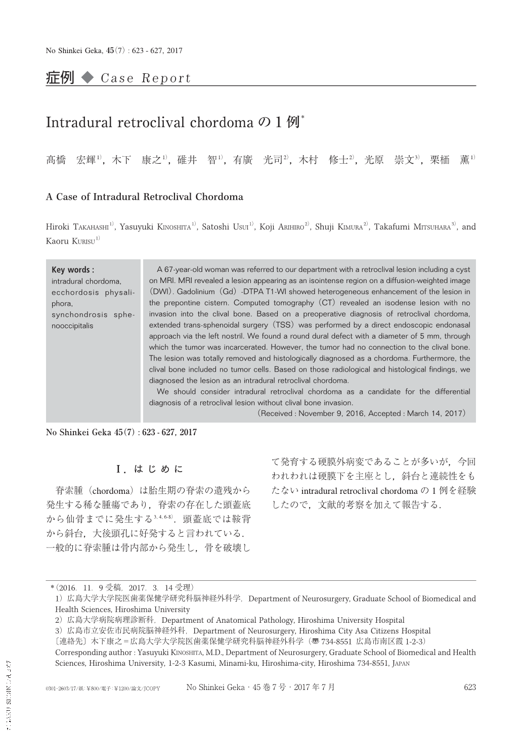
Ⅰ.はじめに
脊索腫(chordoma)は胎生期の脊索の遺残から発生する稀な腫瘍であり,脊索の存在した頭蓋底から仙骨までに発生する3,4,6-8).頭蓋底では鞍背から斜台,大後頭孔に好発すると言われている.一般的に脊索腫は骨内部から発生し,骨を破壊して発育する硬膜外病変であることが多いが,今回われわれは硬膜下を主座とし,斜台と連続性をもたないintradural retroclival chordomaの1例を経験したので,文献的考察を加えて報告する.
A 67-year-old woman was referred to our department with a retroclival lesion including a cyst on MRI. MRI revealed a lesion appearing as an isointense region on a diffusion-weighted image(DWI). Gadolinium(Gd)-DTPA T1-WI showed heterogeneous enhancement of the lesion in the prepontine cistern. Computed tomography(CT)revealed an isodense lesion with no invasion into the clival bone. Based on a preoperative diagnosis of retroclival chordoma, extended trans-sphenoidal surgery(TSS)was performed by a direct endoscopic endonasal approach via the left nostril. We found a round dural defect with a diameter of 5 mm, through which the tumor was incarcerated. However, the tumor had no connection to the clival bone. The lesion was totally removed and histologically diagnosed as a chordoma. Furthermore, the clival bone included no tumor cells. Based on those radiological and histological findings, we diagnosed the lesion as an intradural retroclival chordoma.
We should consider intradural retroclival chordoma as a candidate for the differential diagnosis of a retroclival lesion without clival bone invasion.
Copyright © 2017, Igaku-Shoin Ltd. All rights reserved.