Japanese
English
- 有料閲覧
PPV購入案内
1,320円 (1,200円+税10%) こちらは、233ページから238ページの文献です。購入には医書.jp本会員登録が必要です。
会員登録完了後に改めて上記「購入サイトへ」を選択してください。
または「購入サイトへ」選択後に「購入ログイン」、「新規会員登録」の順に進んでください。
会員登録について詳しくはをご確認ください。
- Abstract 文献概要
- 1ページ目 Look Inside
- 参考文献 Reference
Ⅰ.はじめに
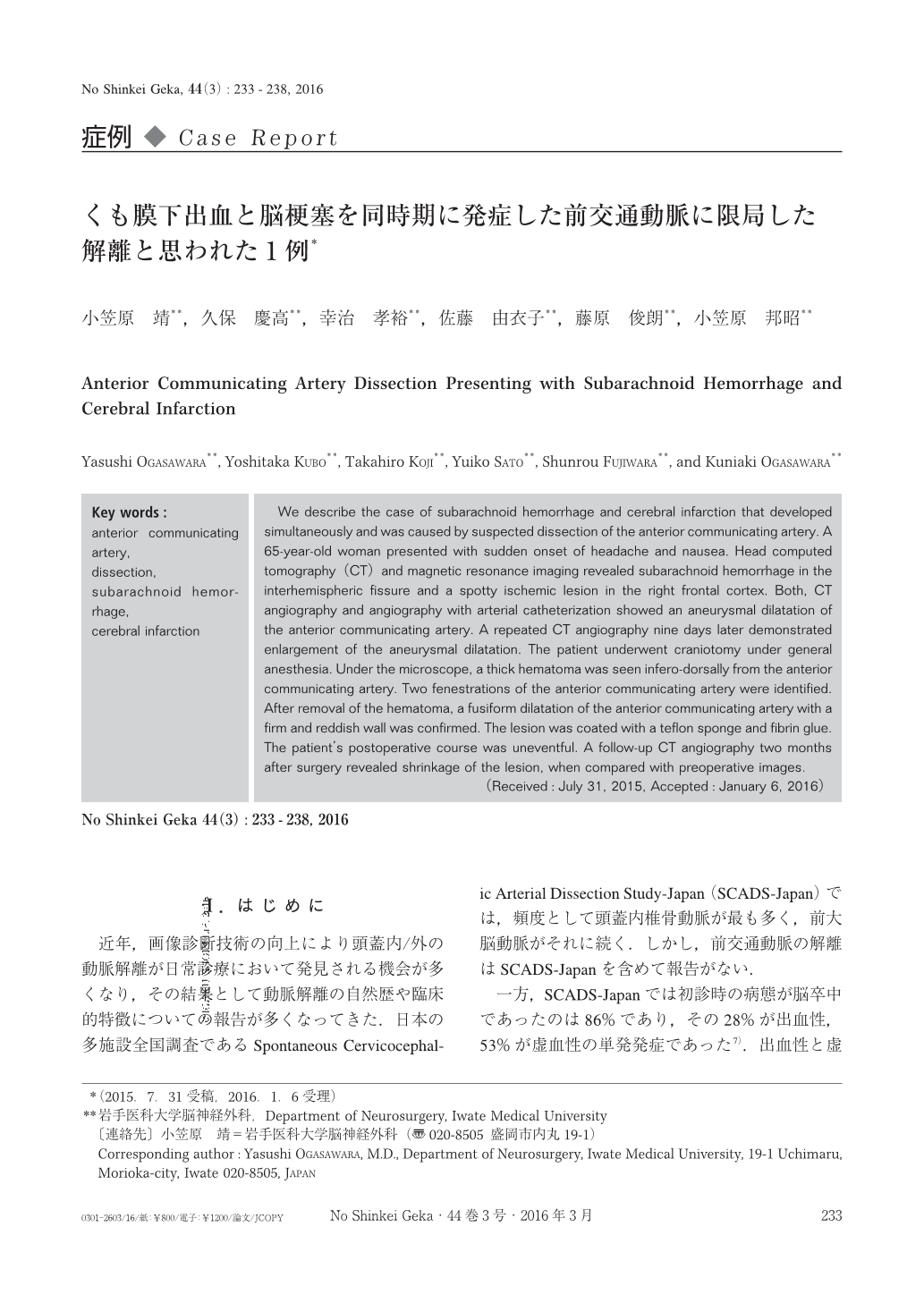
近年,画像診断技術の向上により頭蓋内/外の動脈解離が日常診療において発見される機会が多くなり,その結果として動脈解離の自然歴や臨床的特徴についての報告が多くなってきた.日本の多施設全国調査であるSpontaneous Cervicocephalic Arterial Dissection Study-Japan(SCADS-Japan)では,頻度として頭蓋内椎骨動脈が最も多く,前大脳動脈がそれに続く.しかし,前交通動脈の解離はSCADS-Japanを含めて報告がない.
一方,SCADS-Japanでは初診時の病態が脳卒中であったのは86%であり,その28%が出血性,53%が虚血性の単発発症であった7).出血性と虚血性の脳卒中を同時に認めた症例は5%と少ない7).
今回,われわれはくも膜下出血と脳梗塞を同時に発症し,画像の経時的変化と術中所見から前交通動脈に限局した動脈解離と考えられた1例を経験したので,文献的考察を加えて報告する.
We describe the case of subarachnoid hemorrhage and cerebral infarction that developed simultaneously and was caused by suspected dissection of the anterior communicating artery. A 65-year-old woman presented with sudden onset of headache and nausea. Head computed tomography(CT)and magnetic resonance imaging revealed subarachnoid hemorrhage in the interhemispheric fissure and a spotty ischemic lesion in the right frontal cortex. Both, CT angiography and angiography with arterial catheterization showed an aneurysmal dilatation of the anterior communicating artery. A repeated CT angiography nine days later demonstrated enlargement of the aneurysmal dilatation. The patient underwent craniotomy under general anesthesia. Under the microscope, a thick hematoma was seen infero-dorsally from the anterior communicating artery. Two fenestrations of the anterior communicating artery were identified. After removal of the hematoma, a fusiform dilatation of the anterior communicating artery with a firm and reddish wall was confirmed. The lesion was coated with a teflon sponge and fibrin glue. The patient's postoperative course was uneventful. A follow-up CT angiography two months after surgery revealed shrinkage of the lesion, when compared with preoperative images.
Copyright © 2016, Igaku-Shoin Ltd. All rights reserved.