Japanese
English
- 有料閲覧
PPV購入案内
1,320円 (1,200円+税10%) こちらは、323ページから329ページの文献です。購入には医書.jp本会員登録が必要です。
会員登録完了後に改めて上記「購入サイトへ」を選択してください。
または「購入サイトへ」選択後に「購入ログイン」、「新規会員登録」の順に進んでください。
会員登録について詳しくはをご確認ください。
- Abstract 文献概要
- 1ページ目 Look Inside
- 参考文献 Reference
Ⅰ.はじめに
フラッシュ刺激視覚誘発電位(visual evoked potential:VEP)は,網膜に対する光刺激により大脳皮質視覚野に生じる電位を記録する神経生理検査の1つである.被験者が光刺激画面を意図的に注視する必要がないため,検査への協力が得られない小児例や意識障害患者に対しても施行可能であり4,7,12)1970年代から臨床応用が進められてきたが,安定した電位の記録が困難であることが課題とされてきた21).近年,これらの課題に対して光刺激装置の改良や網膜電図の併用14),完全静脈麻酔の導入6,15,18)が試みられることにより,視覚路近傍の手術の際の術中モニタリングとしてその有用性が再び注目されている3,16).
頭蓋咽頭腫は小児と成人に発生のピークのある良性腫瘍である.腫瘍による視交叉圧迫のために視力視野障害を呈することが多く,頭蓋咽頭腫の手術において視機能の温存や改善は主目的の1つである.手術中に視機能のモニタリングを正確に行うことができれば,手術操作による視覚路の障害が回避可能となるため,術中VEPモニタリングは視機能温存に有用であると期待される.一方で,既に視機能障害を呈している場合,術中モニタリングでその改善がとらえられるかどうかは議論があり,決着をみていない3,11).
今回われわれは,トルコ鞍部頭蓋咽頭腫患者に対して経蝶形骨洞的腫瘍摘出術中にVEPモニタリングを行い,振幅増大と術後臨床症状の改善を認めた症例を経験した.大型のトルコ鞍部腫瘍患者に対して,視機能評価におけるVEPの有用性について文献的考察を加えて報告する.
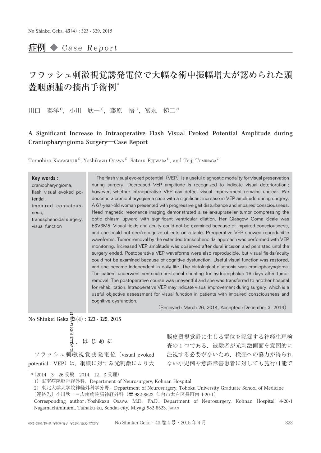
The flash visual evoked potential(VEP)is a useful diagnostic modality for visual preservation during surgery. Decreased VEP amplitude is recognized to indicate visual deterioration;however, whether intraoperative VEP can detect visual improvement remains unclear. We describe a craniopharyngioma case with a significant increase in VEP amplitude during surgery. A 67-year-old woman presented with progressive gait disturbance and impaired consciousness. Head magnetic resonance imaging demonstrated a sellar-suprasellar tumor compressing the optic chiasm upward with significant ventricular dilation. Her Glasgow Coma Scale was E3V3M5. Visual fields and acuity could not be examined because of impaired consciousness, and she could not see/recognize objects on a table. Preoperative VEP showed reproducible waveforms. Tumor removal by the extended transsphenoidal approach was performed with VEP monitoring. Increased VEP amplitude was observed after dural incision and persisted until the surgery ended. Postoperative VEP waveforms were also reproducible, but visual fields/acuity could not be examined because of cognitive dysfunction. Useful visual function was restored, and she became independent in daily life. The histological diagnosis was craniopharyngioma. The patient underwent ventriculo-peritoneal shunting for hydrocephalus 16 days after tumor removal. The postoperative course was uneventful and she was transferred to another hospital for rehabilitation. Intraoperative VEP may indicate visual improvement during surgery, which is a useful objective assessment for visual function in patients with impaired consciousness and cognitive dysfunction.
Copyright © 2015, Igaku-Shoin Ltd. All rights reserved.