Japanese
English
- 有料閲覧
PPV購入案内
1,320円 (1,200円+税10%) こちらは、275ページから280ページの文献です。購入には医書.jp本会員登録が必要です。
会員登録完了後に改めて上記「購入サイトへ」を選択してください。
または「購入サイトへ」選択後に「購入ログイン」、「新規会員登録」の順に進んでください。
会員登録について詳しくはをご確認ください。
- Abstract 文献概要
- 1ページ目 Look Inside
I.はじめに
細菌性脳動脈瘤は極めて短期間に形成され,その形態は短期間に変化することが知られている4-6,8,9,13,15-20,22).また,周辺血管では,攣縮,狭窄,閉塞などの所見も動脈瘤と同時に観察されている場合もある1,2,5,8,9,11,13,15-20,22).それらの周辺血管の変化については,一般にくも膜下出血においてみられる脳血管攣縮と同様の血管攣縮,あるいは血管炎による狭窄,細菌塞栓による閉塞,感染に伴う脳血管攣縮などが考えられるが,充分な検討はいまだなされていない.
今回われわれは,心内膜炎からの細菌性脳動脈瘤の破裂後,動脈瘤の形状の変化のみならず,周辺血管の2度にわたる狭窄と寛解を観察し得た症例を経験した.細菌性脳動脈瘤における周辺血管の変化を中心に若干の考察を加えて報告する.
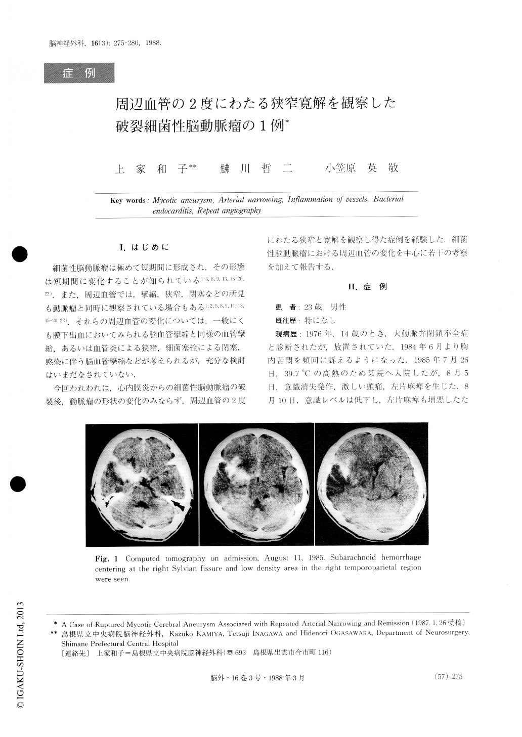
The patient is a 23-year-old male in whom aortic valve regurgitation was pointed out at the age of about 13. From July 20, 1985 he developed a high fever of 39.7℃ and on August 5 he suddenly became uncon-scious with left hemiparesis. On August 10, his con-sciousness became further disturbed and he was admit-ted to this Department on the following day. Computed tomographic scan showed subarachnoid hemorrhageand a low density area in the right temporoparietal lobe.
Copyright © 1988, Igaku-Shoin Ltd. All rights reserved.