Japanese
English
- 有料閲覧
- Abstract 文献概要
- 1ページ目 Look Inside
I.はじめに
椎骨脳底動脈系に生ずる動脈瘤は全頭蓋内動脈瘤の約10%前後で,その好発部位は脳底動脈分岐部,次いで後下小脳動脈分岐部であり,後下小脳動脈末梢部に発生する動脈瘤は1%以下と稀である.今回,われわれは2例の後下小脳動脈末梢部動脈瘤を経験したので,主に診断上のpitfallについて若干の文献的考察を加えて報告する.
Two cases treating aneurysms of the distal PICA were reported, and 36 cases with 39 aneurysms in the literatures in Japan were reviewed concerning the dis. tribution of aneurysms and their findings on CT.
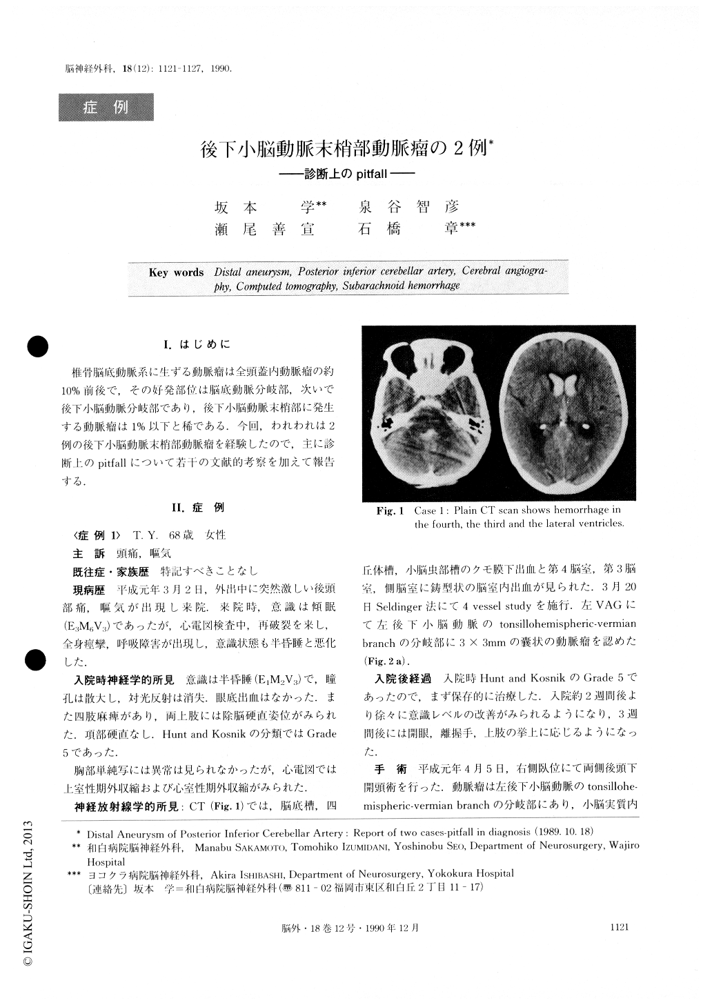
Case 1 ; a 68-year-old female suffered from sudden onset of severe headache and nausea. On admission, it was found she was lethargic. However, her conscious ness deteriorated down to semicoma with tetraparetic condition soon after. CT revealed subarachnoid hemor-rhage in the basal, quadrigeminal and supravermian cisterns and blood clots in the entire ventricle. Cerebral angiography demonstrated an aneurysm located at the distal segment of the left PICA.
She was initially treated conservatively because of being in Hunt and Kosnik Grade 5, and then, 3 weeks after onset, suboccipital craniectomy was performed and the aneurysm was clipped successfully.
Case 2 ; a 60-year-old, female, suddenly experienced severe suboccipitalgia and vomitting. CT revealed sub-arachnoid hemorrhage in the entire subarachnoid space and intraventricular hemorrhages in the 4th, 3rd and lateral ventricles. Subsequently cerebral angiography was performed and left VAG demonstrated an aneurysm at the left A2_-A3 junction. She underwent bi-frontal craniotomy and the aneurysm was clipped via the interhemispheric approach.
Her postoperative course was uneventful. Postopera-tive left CAG showed successful clipping of the aneurysm. However, left VAG suggested an aneurysm-like shadow in the right PICA. Right BAG carried out one week later demonstrated an aneurysm at the distal segment of the right PICA. This aneurysm was then clipped successfully under suboccipital craniectomy.
Distal PICA aneurysm are relatively easy to miss in routine cerebral angiography unless there is adequate reflux of the contrast material on a unilateral VAG to fill the contralateral vertebral artery to the distal seg-ment of the PICA.
From our experience, we would like to emphasize the importance of 4 vessel study in all case of subarachnoid hemorrhage. There were 16 patients who had CT with-in 3 days of their ictus. Thirteen (81%) had the evi-dence of blood in the fourth, 9 (56%) in the third and 5 (31%) in the lateral ventricle. In three cases, hemor-rhage was noted in the ventricles but not in the cistern and in one hemorrhage was confined to supravermian cistern, and in another it was confined to the vermian cistern.
We suggest that there is the possibility of rupture of the distal PICA aneurysm in cases of subarachnoid hemorrhage only within the quadrigeminal and/or ver-mian cistern, or in cases of hemorrhage in the fourth ventricle without subarachnoid hemorrhage.
Copyright © 1990, Igaku-Shoin Ltd. All rights reserved.