Japanese
English
- 有料閲覧
PPV購入案内
1,320円 (1,200円+税10%) こちらは、409ページから414ページの文献です。購入には医書.jp本会員登録が必要です。
会員登録完了後に改めて上記「購入サイトへ」を選択してください。
または「購入サイトへ」選択後に「購入ログイン」、「新規会員登録」の順に進んでください。
会員登録について詳しくはをご確認ください。
- Abstract 文献概要
- 1ページ目 Look Inside
I.緒言
視神経膠腫は全頭蓋内腫瘍の約1%といわれているが5,11),一側の視神経に限局する例はさらにそのなかの半数以下とされている4,14,17).われわれは最近,CT scanにて一側の視神経に限局する視神経膠腫と診断し,手術的にほぼ全摘しえた2例を経験した.主にCT scanによる視神経膠腫の早期診断および手術適応について,若干の文献的考察を加えて報告する.
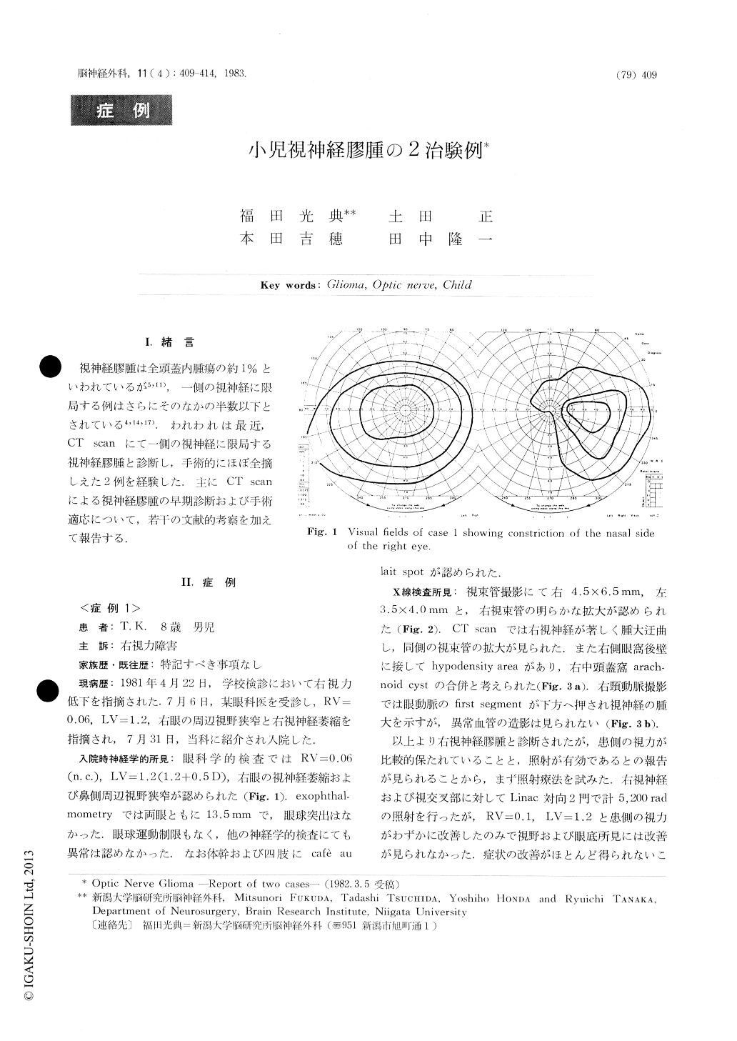
Case 1: 8-year-old boy was admitted to our hospitalcomplaining of right visual impairment. Visualacuity was 0.06 in the right and 1.2 in the left eye, withperipheral constriction in the visual field of the righteye. His right optic fundus showed a simple opticatrophy. There were no proptosis or limitation ofocular movements. Cafe au lait spots were found onthe trunk and extremities as well.
Roentgenograms showed an enlargement of theright optic canal without erosion of sella turcica.Computerized tomography (CT) revealed a markedswelling and tortuosity of the right optic nerve.
Copyright © 1983, Igaku-Shoin Ltd. All rights reserved.