Japanese
English
- 有料閲覧
- Abstract 文献概要
- 1ページ目 Look Inside
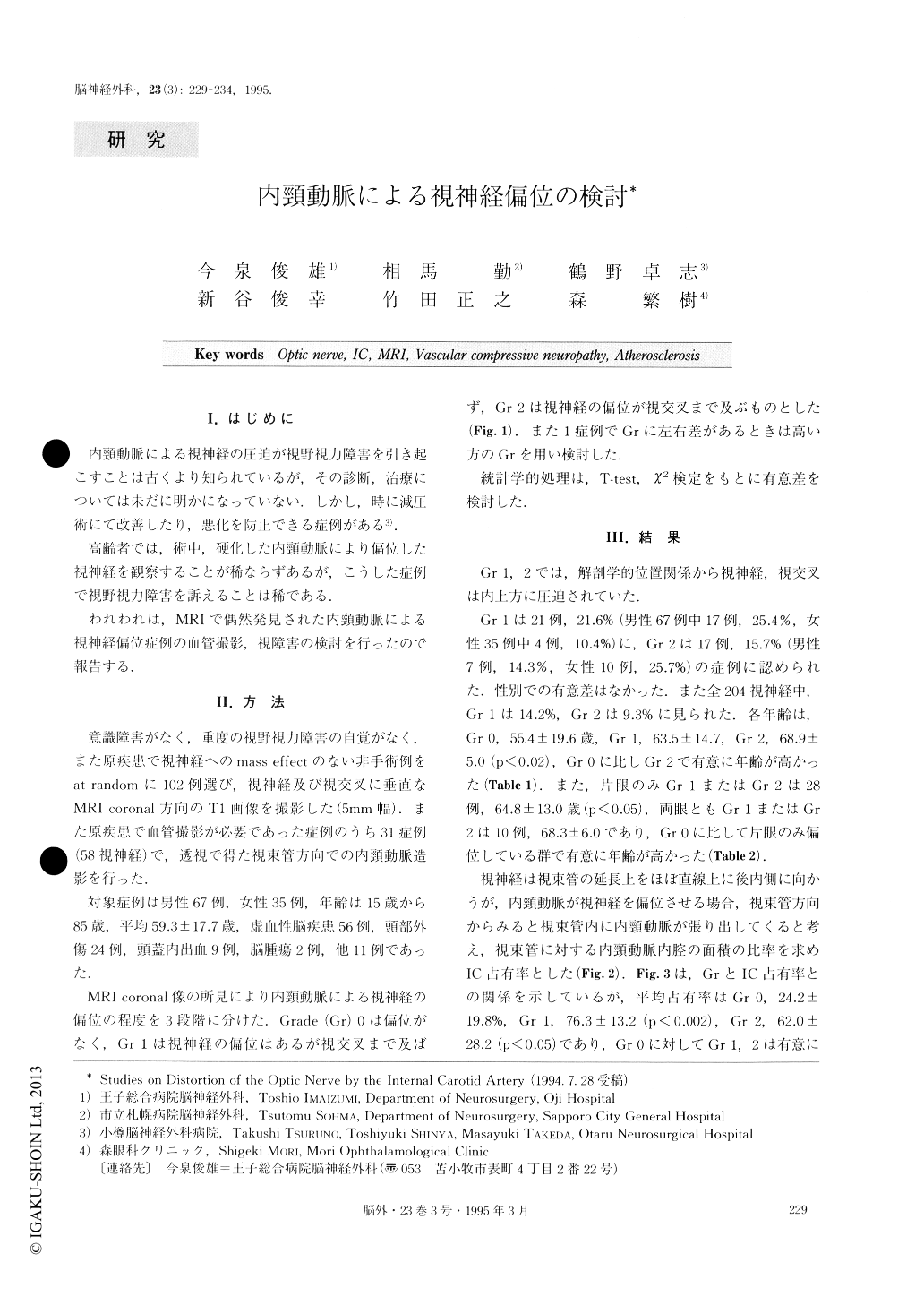
I.はじめに
内頸動脈による視神経の圧迫が視野視力障害を引き起こすことは古くより知られているが,その診断,治療については未だに明かになっていない.しかし,時に減圧術にて改善したり,悪化を防止できる症例がある3).
高齢者では,術中,硬化した内頸動脈により偏位した視神経を観察することが稀ならずあるが,こうした症例で視野視力障害を訴えることは稀である.
The optic nerve (ON) is sometimes distorted by an atherosclerotic internal carotid artery (IC) which we observe during operations. However the distortion sel-dom causes visual dysfunction. In this paper, 102 non-operative cases without severe visual disturbance or marked effect on the visual system were studied, using coronal section of MRI and IC angiography, to ascer-tion ON distortion by IC.
The grading of the distortion was determined by coronal section of MRI. Grade (Gr) 0: No distortion of ON, Gr 1: Distortion of ON without chiasmal disloca-tion, Gr 2 : Distortion of ON with chiasmal dislocation. The rate of Gr 0, Gr 1, and Gr 2 were 62.7, 21.6 and 15.7%, respectively.
The age of Gr 0, Gr 1 and Gr 2 were 55.4 ± 19.6, 63.5 ± 14.7 and 68.9± 5.0 years old, re-spectively (Gr 2 was different from Gr 0). The occupy-ing rate was calculated by the following formula. The areas were measured by IC angiography of the optic canal:
The area of IC in optic canal/The area of optic canal× 100% The rate of Gr 0, Gr 1 and Gr 2 were 24.2 ± 19.8, 76.3 ± 13.2 and 62.0 ± 28.2%, respectively (Gr 1 and 2 differed from Gr 0) . With a rate of more than 70%, the patient had ON distortion by IC. On the antero-posterior views of IC angiography of 1.7 magnifica-tions, the distance from IC to midline of Gr 0, Gr 1 and Gr 2 were 13.5 ± 3.4, 10.7 ± 3.7 and 9.4 ± 2.9mm, re-spectively (Gr 2 differed from Gr 0) . The rate of coil-ing and kinking of cervical IC of Gr 0, Gr 1 and Gr 2 was 13.0, 20.0 and 42.8%, respectively. The distance from the base of the sella to the chiasm of Gr 0, Gr 1 and Gr 2 had no difference. These showed that the atherosclerotic elongation of IC caused the distortion of ON.
The rate of visual disturbance of Gr 0, Gr 1 and Gr 2 was 4.7, 9.1 and 12.5%, respectively. The patients with Gr 2 ON distortion were examined ophthalmologically. Half of them had visual abnormalities which were dia-gnosed as having been caused by cataract, diabetic re-tinopathy, and glaucoma.
Many cases have asymptomatic ON distortion. The distortion seldom makes visual dysfunction. Vascular compressive neuropathy of ON should not be dia-gnosed simply by the radiological finding of ON dis-location.
Copyright © 1995, Igaku-Shoin Ltd. All rights reserved.