Japanese
English
- 有料閲覧
PPV購入案内
1,320円 (1,200円+税10%) こちらは、1005ページから1012ページの文献です。購入には医書.jp本会員登録が必要です。
会員登録完了後に改めて上記「購入サイトへ」を選択してください。
または「購入サイトへ」選択後に「購入ログイン」、「新規会員登録」の順に進んでください。
会員登録について詳しくはをご確認ください。
- Abstract 文献概要
- 1ページ目 Look Inside
I.はじめに
Moyamoya病に対して,近年,精力的に脳血管撮影が施行されるようになってから,脳動脈瘤を合併する症例の報告が増加しつつある.これらの脳動脈瘤の報告を検討してみると,①主幹脳動脈分岐部に発生する通常の動脈瘤(以下,major artery aneurysmという)と,②末梢脳動脈の末端部に発生する動脈瘤(以下,peripheral artery aneurysmという)の2グループに分けられるようであり,この2群の発生機序は異なるように思える.
最近われわれは,前脈絡動脈の末梢部に発生した仮性脳動脈瘤を合併するMoyamoya病を経験したので報告し,文献的考察を行い,Moyamoya病における脳動脈瘤の発生機序について検討を加える.
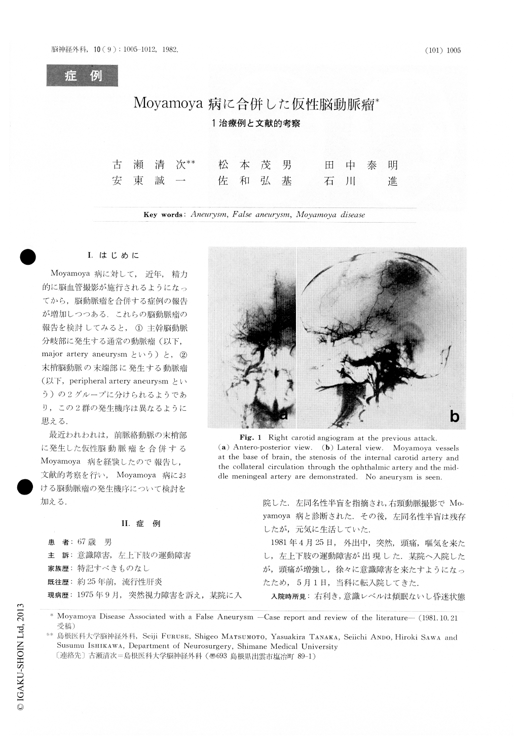
A 67-year-old male was admitted to our clinic on May 1,1981, because of mild confusion and a left hemiparesis.About 6 years ago, when he had a left-sided homonymoushemianopia, carotid angiography revealed moyamoya disease.
CT scan on the admission showed an intracerebralhematoma in the right temporal lobe, which ruptured tothe lateral ventricle. An angiogram demonstrated thenet work of moyamoya vessels in the base of the brain and a"newly-appeared" aneurysm at the peripheral portion ofthe right anterior choroidal artery. This artery was shownon the previous angiogram also but did not bear the aneurysm.
Copyright © 1982, Igaku-Shoin Ltd. All rights reserved.