Japanese
English
- 有料閲覧
PPV購入案内
1,320円 (1,200円+税10%) こちらは、807ページから817ページの文献です。購入には医書.jp本会員登録が必要です。
会員登録完了後に改めて上記「購入サイトへ」を選択してください。
または「購入サイトへ」選択後に「購入ログイン」、「新規会員登録」の順に進んでください。
会員登録について詳しくはをご確認ください。
- Abstract 文献概要
- 1ページ目 Look Inside
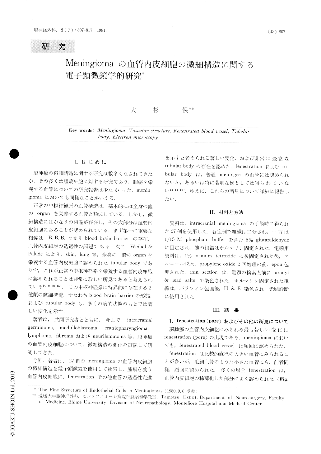
I.はじめに
脳腫瘍の微細構造に関する研究は数多くなされてきたが,その多くは腫瘍細胞に対する研究であり,腫瘍を栄養する血管についての研究報告は少なかった.meningiomaにおいても同様なことがいえる.
正常の中枢神経系の血管構造は,基本的には全身の他のorganを栄養する血管と類似している.しかし,微細構造にはかなりの相違が存在し,その大部分は血管内皮細胞にあることが認められている.まず第一に重要な相違は,B.B.B.つまりblood brain barrierの存在,血管内皮細胞の透過性の問題である.次に,Weibel & Paladeにより,skin, lung等,全身の一般のorganを栄養する血管内皮細胞に認められたtubular bodyであり40),これが正常の中枢神経系を栄養する血管内皮細胞に認められることは非常に珍しい所見であると考えられている9,10,12,15).この中枢神経系に特異的に存在する2種類の微細構造,すなわちblood brain barrierの形態,およびtubular bodyも,多くの病的状態のもとでは著しい変化を示す.
The fine structure of the endothelial cells in meningiomas was studied by the electron microscopy.
There were an increased number of pinocytotic vesicles and fenestrations especially at the attenuated portion of the endothelial cells. Intraluminal infoldings of the plasma membrane were frequently found. Those were certainly abnormal and all probably related to the increased vascular permeability of the endothelial cells.
Copyright © 1981, Igaku-Shoin Ltd. All rights reserved.