Japanese
English
- 有料閲覧
PPV購入案内
1,320円 (1,200円+税10%) こちらは、1445ページから1450ページの文献です。購入には医書.jp本会員登録が必要です。
会員登録完了後に改めて上記「購入サイトへ」を選択してください。
または「購入サイトへ」選択後に「購入ログイン」、「新規会員登録」の順に進んでください。
会員登録について詳しくはをご確認ください。
- Abstract 文献概要
- 1ページ目 Look Inside
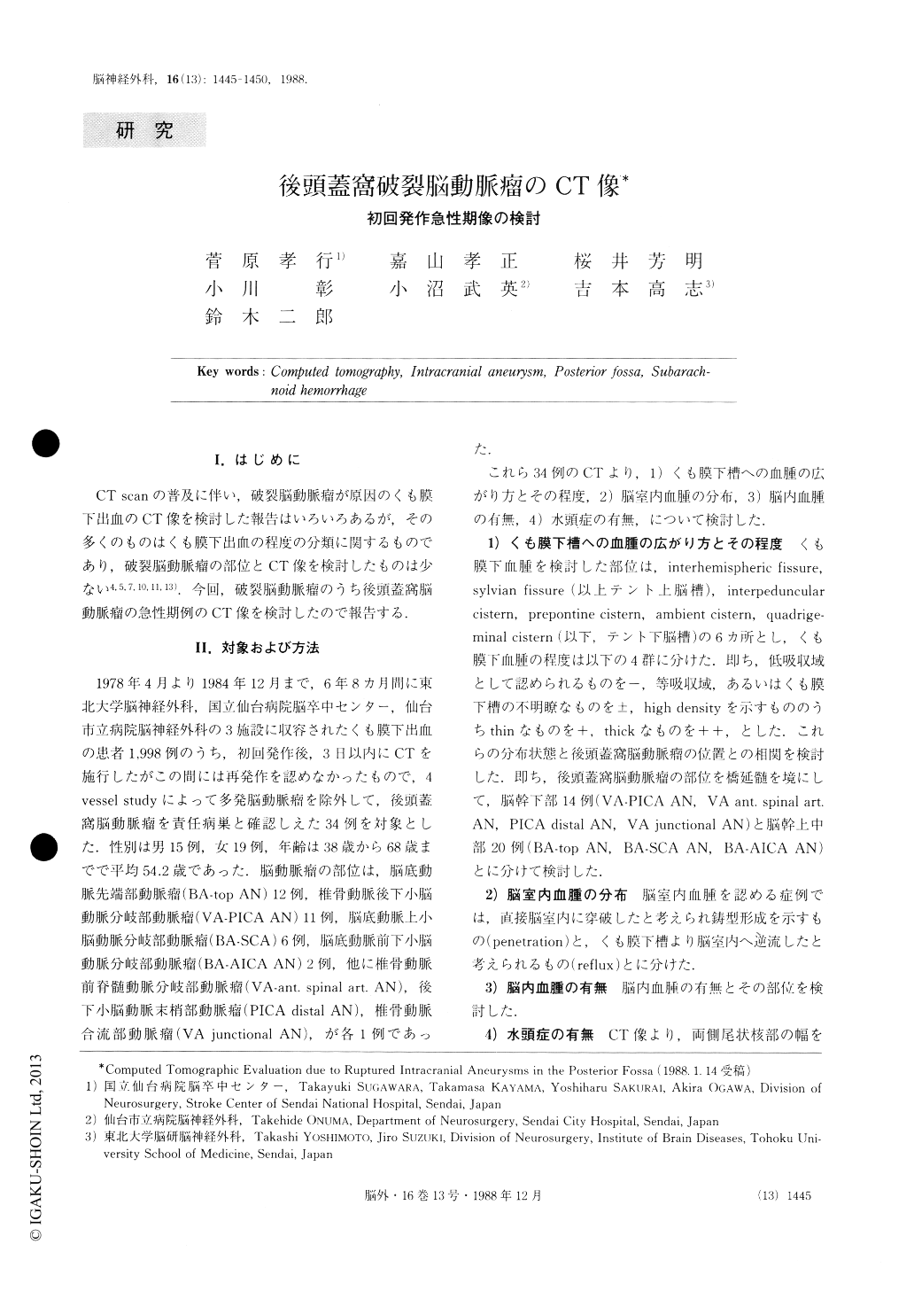
I.はじめに
CT scanの普及に伴い,破裂脳動脈瘤が原因のくも膜下出血のCT像を検討した報告はいろいろあるが,その多くのものはくも膜下出血の程度の分類に関するものであり,破裂脳動脈瘤の部位とCT像を検討したものは少ない4,5,7,10,11,13).今回,破裂脳動脈瘤のうち後頭蓋窩脳動脈瘤の急性期例のCT像を検討したので報告する.
From April, 1978 through December, 1984, computed tomographic (CT) findings were carefully examined in 34 cases of initial subarachnoid bleeding due to a single ruptured aneurysm in the posterior fossa. All of the pa-tients were hospitalized within 3 days of the onset of symptoms. High-density areas, which indicate the pre-sence of subarachnoid clots, were evaluated in the in-terhemispheric and Sylvian fissures and the interpe-duncular, prepontine, ambient, and quadrigeminal cist-erns. The CT data suggest that hematomas in the four cisterns are thicker than those in the supratentorial sub-arachnoid spaces.
Copyright © 1988, Igaku-Shoin Ltd. All rights reserved.