Japanese
English
- 有料閲覧
PPV購入案内
1,320円 (1,200円+税10%) こちらは、1051ページから1056ページの文献です。購入には医書.jp本会員登録が必要です。
会員登録完了後に改めて上記「購入サイトへ」を選択してください。
または「購入サイトへ」選択後に「購入ログイン」、「新規会員登録」の順に進んでください。
会員登録について詳しくはをご確認ください。
- Abstract 文献概要
- 1ページ目 Look Inside
- 参考文献 Reference
Ⅰ.はじめに
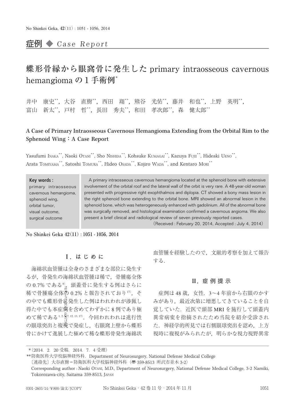
海綿状血管腫は全身のさまざまな部位に発生するが,骨発生の海綿状血管腫は稀で,骨腫瘍全体の0.7%である9).頭蓋骨に発生する例はさらに稀で骨腫瘍全体の0.2%と報告されており17),その中でも蝶形骨に発生した例はわれわれが渉猟し得た中でも本症例を含めてわずかに8例であり極めて稀である1,2,5,7,12,13,17).今回われわれは進行性の眼球突出と複視で発症し,右眼窩上壁から蝶形骨にかけて進展した極めて稀な蝶形骨発生海綿状血管腫を経験したので,文献的考察を加えて報告する.
A primary intraosseous cavernous hemangioma located at the sphenoid bone with extensive involvement of the orbital roof and the lateral wall of the orbit is very rare. A 48-year-old woman presented with progressive right exophthalmos and diplopia. CT showed a bony mass lesion in the right sphenoid bone extending to the orbital bone. MRI showed an abnormal lesion in the sphenoid bone, which was heterogeneously enhanced with gadolinium. All of the abnormal bone was surgically removed, and histological examination confirmed a cavernous angioma. We also present a brief clinical and radiological review of seven previously reported cases.
Copyright © 2014, Igaku-Shoin Ltd. All rights reserved.