Japanese
English
- 有料閲覧
PPV購入案内
1,320円 (1,200円+税10%) こちらは、341ページから347ページの文献です。購入には医書.jp本会員登録が必要です。
会員登録完了後に改めて上記「購入サイトへ」を選択してください。
または「購入サイトへ」選択後に「購入ログイン」、「新規会員登録」の順に進んでください。
会員登録について詳しくはをご確認ください。
- Abstract 文献概要
- 1ページ目 Look Inside
- 参考文献 Reference
Ⅰ.はじめに
悪性黒色腫は,脳実質,軟膜,粘膜組織および皮膚に存在するメラノサイトに由来する悪性腫瘍である.中枢神経原発悪性黒色腫(primary intracranial malignant melanoma:PIMM)は,悪性黒色腫の中でも約1%と極めて稀であり,原発性脳腫瘍の中でも0.1%とされる3,6).初期診断時に,頭蓋内圧亢進(43%),神経学的脱落徴候(35%),痙攣もしくはくも膜下出血(16%)を呈し発見されることが多い6).発見されてからの生存期間中央値は12カ月とされ4),極めて予後不良な疾患である.今回,頭部外傷時に痙攣発作を呈し,偶発的に指摘され当初脳挫傷が疑われていたが,病変の進展後に開頭術により診断されたPIMMの1例について,文献的考察を加えて報告する.
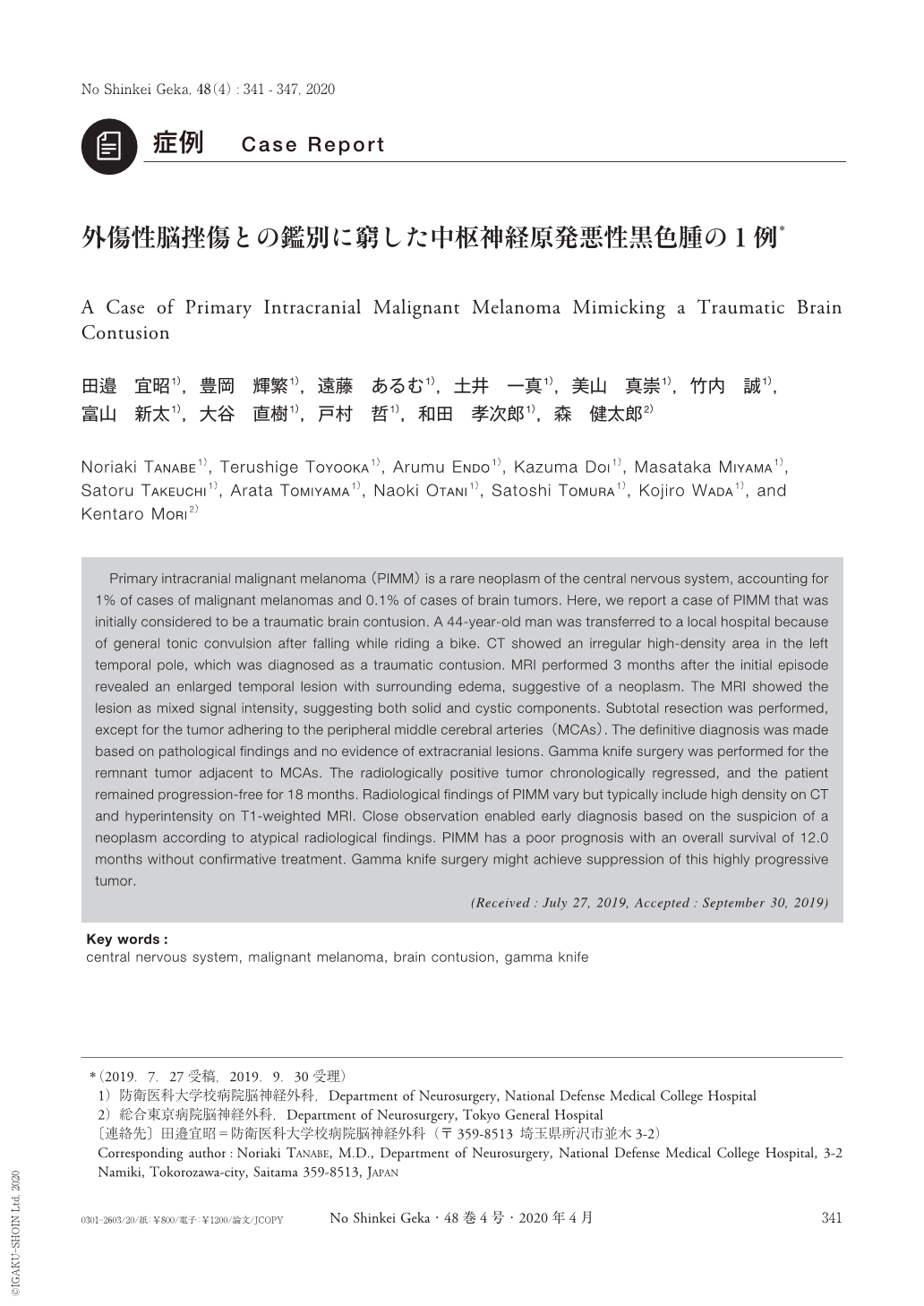
Primary intracranial malignant melanoma(PIMM)is a rare neoplasm of the central nervous system, accounting for 1% of cases of malignant melanomas and 0.1% of cases of brain tumors. Here, we report a case of PIMM that was initially considered to be a traumatic brain contusion. A 44-year-old man was transferred to a local hospital because of general tonic convulsion after falling while riding a bike. CT showed an irregular high-density area in the left temporal pole, which was diagnosed as a traumatic contusion. MRI performed 3 months after the initial episode revealed an enlarged temporal lesion with surrounding edema, suggestive of a neoplasm. The MRI showed the lesion as mixed signal intensity, suggesting both solid and cystic components. Subtotal resection was performed, except for the tumor adhering to the peripheral middle cerebral arteries(MCAs). The definitive diagnosis was made based on pathological findings and no evidence of extracranial lesions. Gamma knife surgery was performed for the remnant tumor adjacent to MCAs. The radiologically positive tumor chronologically regressed, and the patient remained progression-free for 18 months. Radiological findings of PIMM vary but typically include high density on CT and hyperintensity on T1-weighted MRI. Close observation enabled early diagnosis based on the suspicion of a neoplasm according to atypical radiological findings. PIMM has a poor prognosis with an overall survival of 12.0 months without confirmative treatment. Gamma knife surgery might achieve suppression of this highly progressive tumor.
Copyright © 2020, Igaku-Shoin Ltd. All rights reserved.