Japanese
English
- 有料閲覧
PPV購入案内
1,320円 (1,200円+税10%) こちらは、421ページから428ページの文献です。購入には医書.jp本会員登録が必要です。
会員登録完了後に改めて上記「購入サイトへ」を選択してください。
または「購入サイトへ」選択後に「購入ログイン」、「新規会員登録」の順に進んでください。
会員登録について詳しくはをご確認ください。
- Abstract 文献概要
- 1ページ目 Look Inside
- 参考文献 Reference
Ⅰ.はじめに
結節性硬化症(tuberous sclerosis)は高頻度にてんかんを合併し,その多くは薬物治療に抵抗性である.しかし,結節が多発している場合はてんかん焦点の同定が困難なため,外科治療が適応できないことも多い.てんかん原性結節の同定にはMRIに加えて,脳磁図(MEG)やFDG-PETなどが用いられるが,特に乳幼児期のてんかんは,全般性の脳波異常や局在所見に乏しいてんかん発作症状を呈する傾向が強く,てんかん焦点の同定は一般に困難である3,9,16).
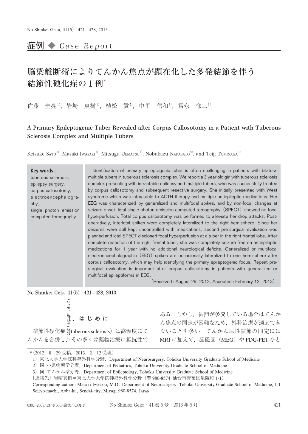
今回われわれは,多発結節を伴う結節性硬化症の小児に対して脳梁離断術を行ったところ,てんかん焦点が明らかになり,二期的にてんかん原性結節を切除して発作の寛解を得た1例を経験したので報告する.
Identification of primary epileptogenic tuber is often challenging in patients with bilateral multiple tubers in tuberous sclerosis complex. We report a 3 year old girl with tuberous sclerosis complex presenting with intractable epilepsy and multiple tubers, who was successfully treated by corpus callosotomy and subsequent resective surgery. She initially presented with West syndrome which was intractable to ACTH therapy and multiple antiepileptic medications. Her EEG was characterized by generalized and multifocal spikes, and by non-focal changes at seizure onset. Ictal single photon emission computed tomography(SPECT)showed no focal hyperperfusion. Total corpus callosotomy was performed to alleviate her drop attacks. Post-operatively, interictal spikes were completely lateralized to the right hemisphere. Since her seizures were still kept uncontrolled with medications, second pre-surgical evaluation was planned and ictal SPECT disclosed focal hyperperfusion at a tuber in the right frontal lobe. After complete resection of the right frontal tuber, she was completely seizure free on antiepileptic medications for 1 year with no additional neurological deficits. Generalized or multifocal electroencephalographic(EEG)spikes are occasionally lateralized to one hemisphere after corpus callosotomy, which may help identifying the primary epileptogenic focus. Repeat pre-surgical evaluation is important after corpus callosotomy in patients with generalized or multifocal epileptiforms in EEG.
Copyright © 2013, Igaku-Shoin Ltd. All rights reserved.