Japanese
English
- 有料閲覧
PPV購入案内
こちらは、991ページから997ページの文献です。購入には医書.jp本会員登録が必要です。
会員登録完了後に改めて上記「購入サイトへ」を選択してください。
または「購入サイトへ」選択後に「購入ログイン」、「新規会員登録」の順に進んでください。
会員登録について詳しくはをご確認ください。
- Abstract 文献概要
- 1ページ目 Look Inside
- 参考文献 Reference
Ⅰ.はじめに
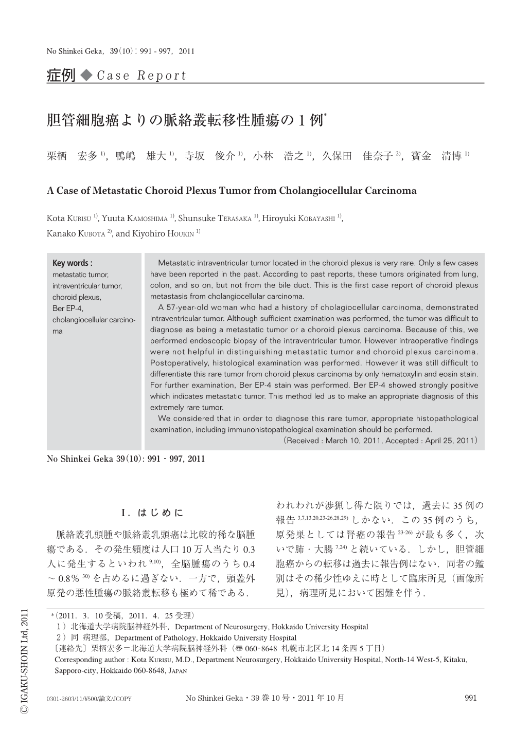
脈絡叢乳頭腫や脈絡叢乳頭癌は比較的稀な脳腫瘍である.その発生頻度は人口10万人当たり0.3人に発生するといわれ9,10),全脳腫瘍のうち0.4~0.8%30)を占めるに過ぎない.一方で,頭蓋外原発の悪性腫瘍の脈絡叢転移も極めて稀である.われわれが渉猟し得た限りでは,過去に35例の報告3,7,13,20,23-26,28,29)しかない.この35例のうち,原発巣としては腎癌の報告23-26)が最も多く,次いで肺・大腸7,24)と続いている.しかし,胆管細胞癌からの転移は過去に報告例はない.両者の鑑別はその稀少性ゆえに時として臨床所見(画像所見),病理所見において困難を伴う.
今回,われわれは成人担癌患者(胆管細胞癌)に側脳室内脈絡叢部に腫瘍が認められた症例を経験した.画像所見,病理所見を含め原発性腫瘍,転移性腫瘍の鑑別が問題となったが,病理組織診断を用いて最終診断として転移性腫瘍との診断に至った.この非常に稀であり,診断に難渋した症例をその診断過程も含めて若干の文献的考察を加えて報告する.
Metastatic intraventricular tumor located in the choroid plexus is very rare. Only a few cases have been reported in the past. According to past reports,these tumors originated from lung,colon,and so on,but not from the bile duct. This is the first case report of choroid plexus metastasis from cholangiocellular carcinoma.
A 57-year-old woman who had a history of cholagiocellular carcinoma, demonstrated intraventricular tumor. Although sufficient examination was performed, the tumor was difficult to diagnose as being a metastatic tumor or a choroid plexus carcinoma. Because of this, we performed endoscopic biopsy of the intraventricular tumor. However intraoperative findings were not helpful in distinguishing metastatic tumor and choroid plexus carcinoma. Postoperatively, histological examination was performed. However it was still difficult to differentiate this rare tumor from choroid plexus carcinoma by only hematoxylin and eosin stain. For further examination, Ber EP-4 stain was performed. Ber EP-4 showed strongly positive which indicates metastatic tumor. This method led us to make an appropriate diagnosis of this extremely rare tumor.
We considered that in order to diagnose this rare tumor,appropriate histopathological examination,including immunohistopathological examination should be performed.
Copyright © 2011, Igaku-Shoin Ltd. All rights reserved.