Japanese
English
- 有料閲覧
PPV購入案内
1,320円 (1,200円+税10%) こちらは、108ページから113ページの文献です。購入には医書.jp本会員登録が必要です。
会員登録完了後に改めて上記「購入サイトへ」を選択してください。
または「購入サイトへ」選択後に「購入ログイン」、「新規会員登録」の順に進んでください。
会員登録について詳しくはをご確認ください。
- Abstract 文献概要
- 1ページ目 Look Inside
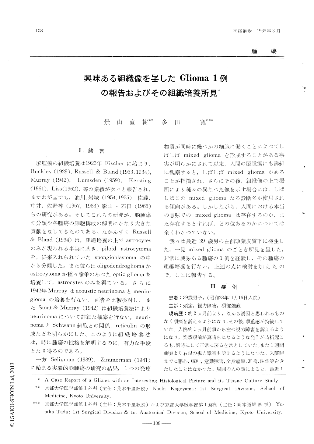
I.緒言
脳腫瘍の組織培養は1925年Fischerに始まり,Buckley(1929),Russell & Bland(1933,1934),Murray(1942),Lumsden(1959),Kersting(1961),Liss(1962),等の業績が次々と報告され,またわが国でも,油川,岩城(1954,1955),佐藤,中井,佐野等(1957,1963)影山・石田(1965)らの研究がある。そしてこれらの研究が,脳腫瘍の分類や各腫瘍の細胞構成の解明にかなり大きな貢献をなしてきたのである。なかんずくRussell & Bland(1934)は,組織培養の上でastmcytesのみが現われる事実に基き,piloid astrocytomaを,従来入れられていたspongioblastomaの中から分離した。また彼らはoligodendrogliomaかastrocytomaか種々論争のあつたoptic gliomaを培養して,astrocytesのみを得ている。さらに1942年Murrayはacoustic neurinomaとmeningiomaの培養を行ない,両者を比較検討し,またStout & Murray(1942)は組織培養法によりneurinomaについて詳細な観察を行ない,neurinomaとSchwann細胞との関係,reticulinの形成などを明らかにした。
A clearly demarcated soft glioma of an egg sizewas surgically excised from the left frontal lobe of a 39-year-old male. Histologically, the tumor was composed of four different areas. 1) The major parts ofthe tumor were composed by groups of glial cellswhich had small, round or oval, chromatin-richnuclei and fine fibrous processes. No necroticareas were observed, but vascular connective tissueshowed hyaline degenerations of high degree (Fig.1-A). 2) Some areas demonstrated astrocytoma-like picture, consisting of multipolar astrocyteswhich were somehow larger than the glial cellsappeared in the foregoing areas. Abundant fine fibres composed the ground of those areas (Fig. 1-B). 3) Other areas consisting of closely packedglial cells with round nuclei and clear cytoplasmresembled the histological picture of oligodendroglioma (Fig. 1-C). 4) Areas of ependymoma-likepicture were also observed, where pseudorosette-like structures were seen at a glance (Fig. 1-D).
Tissue culture of this tumor revealed only twotypes of glial cells to migrate juvenile astrocytes and small undifferentiated glial cells of spindle shape resembling socalled spongioblasts. Noother glial cells were observed to migrate at anystage of the culture (Fig. 2 & 3).
Careful reobservation of the histological pictureof this glioma revealed that above-mentioned oligo-dendrogliomadike or ependymoma-like pictures werenot identical with the genuine histological picturesof typical oligodendroglioma or ependymoma. Thecells in these areas could be considered as variations of astrocytic component. This glioma wastherefore diagnosed as glioblastoma or spongioblastoma. We would like to emphasize the fact thatcells of astrocytic origin, as being described above, will sometimes demonstrate confusious histologicalpictures, but careful observation of the histologicalpicture of the specimen will prevent us from making misdiagnosis.
Copyright © 1965, Igaku-Shoin Ltd. All rights reserved.