Japanese
English
- 有料閲覧
- Abstract 文献概要
- 1ページ目 Look Inside
Ⅰ.はじめに
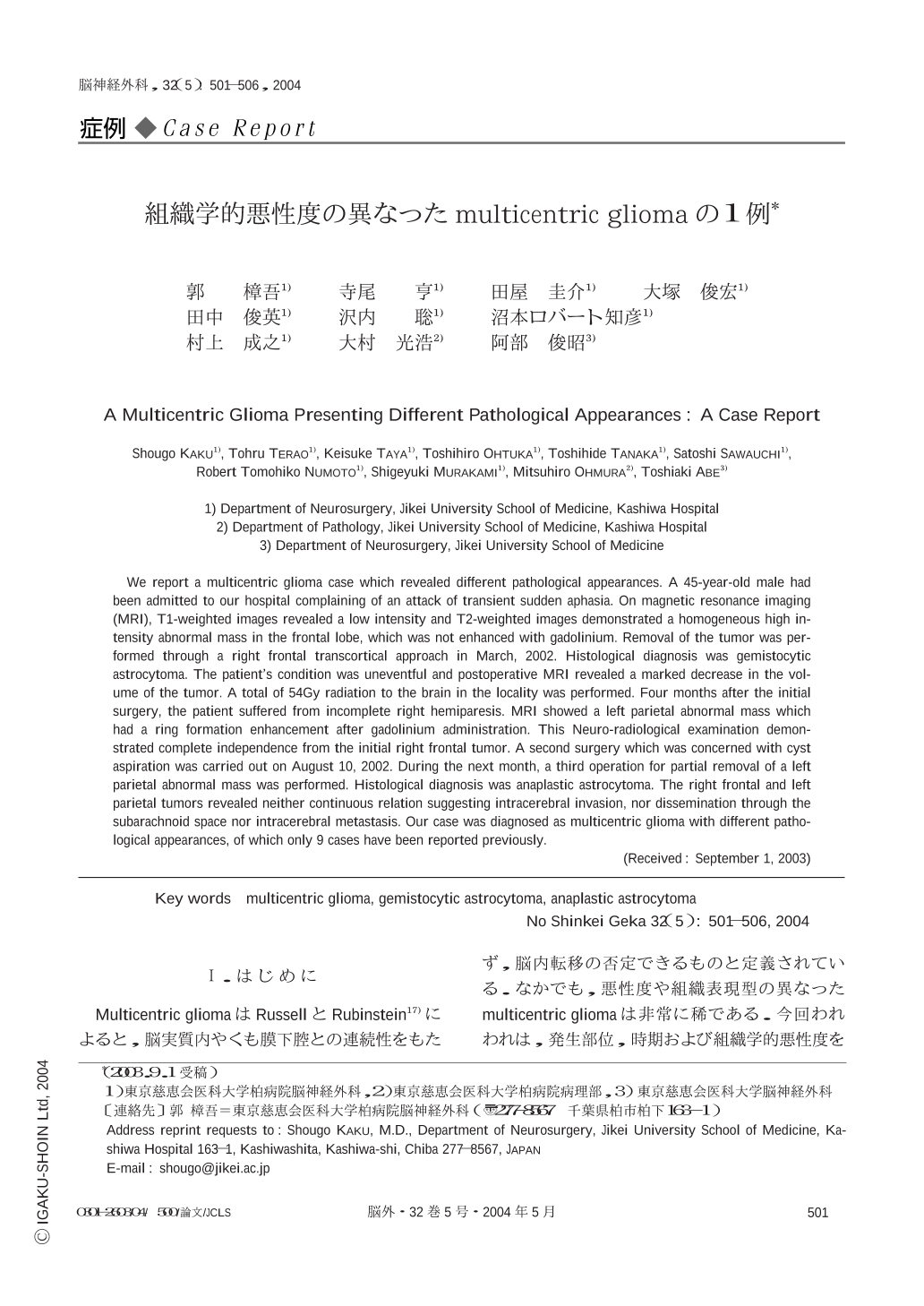
Multicentric gliomaはRussellとRubinstein17)によると,脳実質内やくも膜下腔との連続性をもたず,脳内転移の否定できるものと定義されている.なかでも,悪性度や組織表現型の異なったmulticentric gliomaは非常に稀である.今回われわれは,発生部位,時期および組織学的悪性度を異にする2つのgliomaとして診断されたmulticentric gliomaの1例を経験したので,文献的考察を加え報告する.
We report a multicentric glioma case which revealed different pathological appearances. A 45-year-old male had been admitted to our hospital complaining of an attack of transient sudden aphasia. On magnetic resonance imaging (MRI),T1-weighted images revealed a low intensity and T2-weighted images demonstrated a homogeneous high intensity abnormal mass in the frontal lobe,which was not enhanced with gadolinium. Removal of the tumor was performed through a right frontal transcortical approach in March,2002. Histological diagnosis was gemistocytic astrocytoma. The patient's condition was uneventful and postoperative MRI revealed a marked decrease in the volume of the tumor. A total of 54Gy radiation to the brain in the locality was performed. Four months after the initial surgery,the patient suffered from incomplete right hemiparesis. MRI showed a left parietal abnormal mass which had a ring formation enhancement after gadolinium administration. This Neuro-radiological examination demonstrated complete independence from the initial right frontal tumor. A second surgery which was concerned with cyst aspiration was carried out on August 10,2002. During the next month,a third operation for partial removal of a left parietal abnormal mass was performed. Histological diagnosis was anaplastic astrocytoma. The right frontal and left parietal tumors revealed neither continuous relation suggesting intracerebral invasion,nor dissemination through the subarachnoid space nor intracerebral metastasis. Our case was diagnosed as multicentric glioma with different pathological appearances,of which only 9 cases have been reported previously.

Copyright © 2004, Igaku-Shoin Ltd. All rights reserved.


