Japanese
English
- 有料閲覧
- Abstract 文献概要
- 1ページ目 Look Inside
I.はじめに
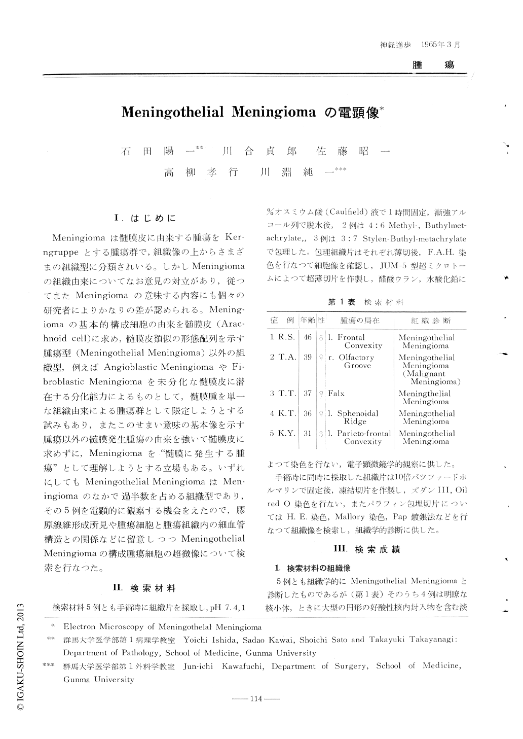
Meningiomaは髄膜皮に由来する腫瘍をKerngruppeとする腫瘍群で,組織像の上からさまざまの組織型に分類されいる。しかしMeningiomaの組織由来についてなお意見の対立があり,従つてまたMeningiomaの意味する内容にも個々の研究者によりかなりの差が認められる。Meningiomaの基本的構成細胞の由来を髄膜皮(Arachnoid cell)に求め,髄膜皮類似の形態配列を示す腫瘍型(Meningothelial Meningioma)の以外の組織型,例えばAngioblastic MeninglomaやFibroblastic Meningiomaを未分化な髄膜皮に潜在する分化能力によるものとして,髄膜腫を単一な組織由来による腫瘍群として限定しようとする試みもあり,またこのせまい意味の基本像を示す腫瘍以外の髄膜発生腫瘍の由来を強いて髄膜皮に求めずに,Meningiomaを"髄膜に発生する腫瘍"として理解しようとする立場もある。いずれにしてもMeningothelial MeningiomaはMeningiomaのなかで過半数を占める組織型であり,その5例を電顕的に観察する機会をえたので,膠原線維形成所見や腫瘍細胞と腫瘍組織内の細血管構造との関係などに留意しつつMeningothelial Meningiomaの構成腫瘍細胞の超微像について検索を行なつた。
Five meningiomas of meningothelial variety wereexamined with electron microscope. "Fite neoplastk-meningothelial cells appeared to he characterizedby their fine cellular details closely resemblingthose of arachnid cells and by complicated surfacespecialization. The nuclei were relatively regularin outline, containing occasionally inclusions. Thecytoplasm were pale and abundant, and most commonly found in the form of long irregular pseudopods or extensions which fit together and interdigitated with those of adjacent cells. They containedonly scant rihonucleoprotein granules and scatteredcytoplasmic organelles including vesicles. Occasional tumors were characterized by the abundanceof fine fibrils in the cytoplasm. The elongated cellswere found in areas encircling together formingcell whorls. In cases desmosomes were prominently evidenced at the margin of the cells. The finestructural features were even demonstrated in atumor classified as malignant type of meningioma.
It is a question of interest whether the rneingioma cells do posess a fibroblastic or angioblasticactivity, In areas collagenous fibers were found. running between the cells. They appeared to blend. with the cell membrane, indicating the possibilityof fibroblastic activity of meningothelial tumor cells. A tumor examined showed a rich net work of vascular and intercellular spaces, which were seenenclosed by tumor cells with prominent attachmentplates. In areas where the light microscopy presented a pseudoxanthomatous appearance, tumor cellcytoplasms were filled with vacuoles, containingitmorphous materials of lipid in nature.

Copyright © 1965, Igaku-Shoin Ltd. All rights reserved.


