Japanese
English
- 有料閲覧
PPV購入案内
1,320円 (1,200円+税10%) こちらは、690ページから692ページの文献です。購入には医書.jp本会員登録が必要です。
会員登録完了後に改めて上記「購入サイトへ」を選択してください。
または「購入サイトへ」選択後に「購入ログイン」、「新規会員登録」の順に進んでください。
会員登録について詳しくはをご確認ください。
- Abstract 文献概要
- 1ページ目 Look Inside
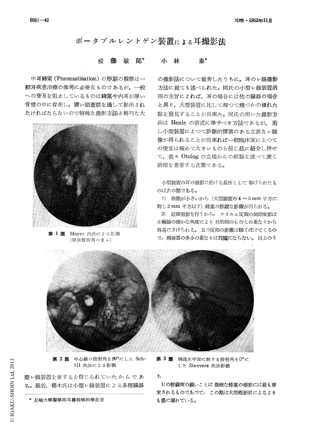
中耳蜂窠(Pneumatisation)の形態の觀察は一般耳疾患治療の參考に必要なものであるが,一般への普及を阻止しているものは蜂窠や内耳が厚い骨壁の中に存在し,廣い頭蓋腔を通して影出されなければならないので特殊な撮影方法と精巧な大型レ線裝置を要すると信じられていたからである。最近,櫻木氏は小型レ線裝置による各種臓器の撮影法について報告したうちに,耳のレ線撮影方法に就ても述べられた。同氏の小型レ線裝置活用の主旨によれば,耳の場合には他の臓器の場合と異り,大型裝置に比して却つて幾つかの優れた點を發見することが出來た。同氏の用いた撮影方向はHenleの術式に準ずべき方法であるが,若し小型裝置によつて診斷的價値のある立派なレ線像が得られることが出來れば一般臨床家にとつての便宜は極めて大きいものと信じ茲に紹介し併せて,我々Otologの立場からの經驗を述べて廣く活用を希望する次第である。
小型裝置の耳の撮影に於ける長所として擧げられたものは次の點である。
GOTO AND KOBAYASHI recommend a method of taking X-ray pictures of the ear with a portable unit that was developed by Sakuragi and recently introduced in literature by the authors. This method not only applies fundamental principles of exposure which was advanced by Schuller, Stevens and, Mayer and special methods of that of Lange and Sonnenkalb but, differing somewhat from such methods, it is noted by having a shorter angle of exposure thereby, giving ease to the procedure with minimum distortion to the picture taken that would be clear-cut in its outlines.
Copyright © 1953, Igaku-Shoin Ltd. All rights reserved.