Japanese
English
- 有料閲覧
PPV購入案内
1,320円 (1,200円+税10%) こちらは、79ページから83ページの文献です。購入には医書.jp本会員登録が必要です。
会員登録完了後に改めて上記「購入サイトへ」を選択してください。
または「購入サイトへ」選択後に「購入ログイン」、「新規会員登録」の順に進んでください。
会員登録について詳しくはをご確認ください。
- Abstract 文献概要
- 1ページ目 Look Inside
- 参考文献 Reference
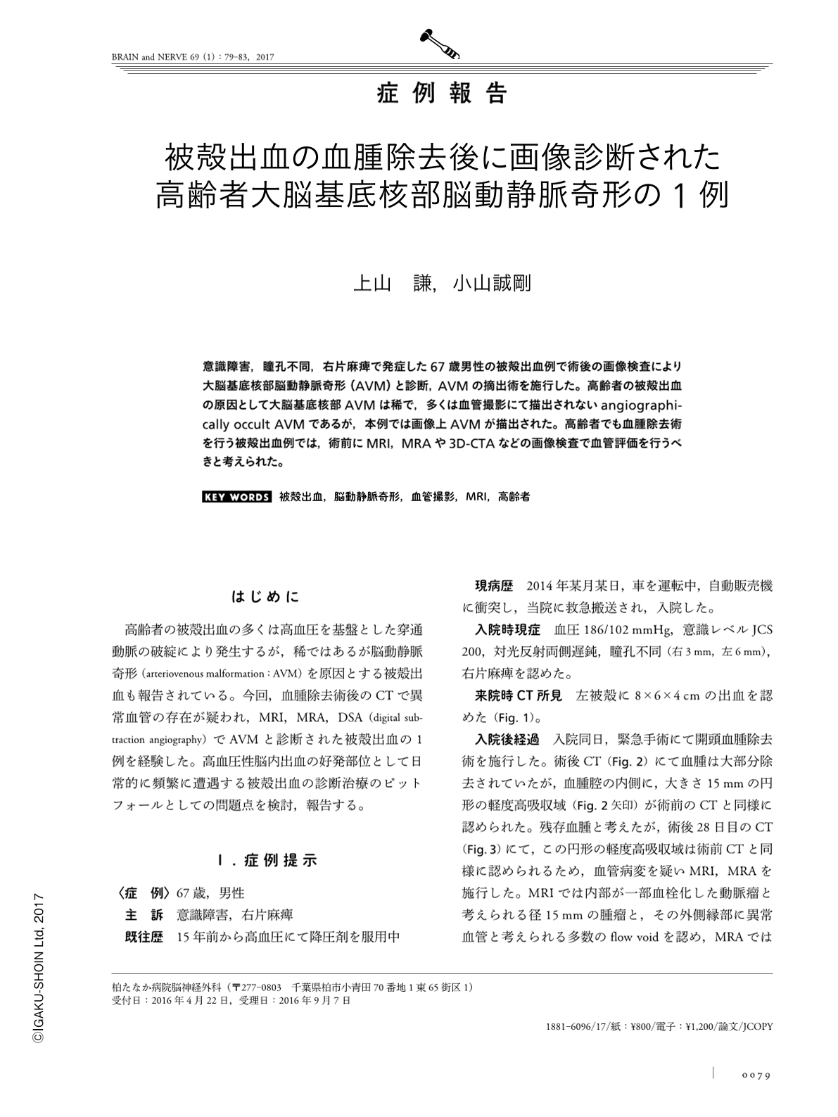
意識障害,瞳孔不同,右片麻痺で発症した67歳男性の被殻出血例で術後の画像検査により大脳基底核部脳動静脈奇形(AVM)と診断,AVMの摘出術を施行した。高齢者の被殻出血の原因として大脳基底核部AVMは稀で,多くは血管撮影にて描出されないangiographically occult AVMであるが,本例では画像上AVMが描出された。高齢者でも血腫除去術を行う被殻出血例では,術前にMRI,MRAや3D-CTAなどの画像検査で血管評価を行うべきと考えられた。
Abstract
A 67-year-old male patient presented with disturbance of consciousness, anisocoria, and right hemiparesis. Computed tomography (CT) scans demonstrated a large putaminal hemorrhage on the left. An emergent operation was performed and the hematoma was removed. Postoperative magnetic resonance angiographic (MRA) images and carotid angiography (CAG) revealed a basal ganglia arteriovenous malformation (AVM). The AVM was resected completely. Putaminal hemorrhages due to basal ganglia AVM rupture are very rare in the elderly. Although most AVMs are angiographically occult, in the current case, the AVM was diagnosed using MRA and CAG. Thus, preoperative examination such as MRA and 3D-CT angiography should be performed in putaminal hemorrhages, even in the elderly.
(Received April 22, 2016; Accepted September 7, 2016; Published January 1, 2017)
Copyright © 2017, Igaku-Shoin Ltd. All rights reserved.