Japanese
English
- 有料閲覧
PPV購入案内
1,320円 (1,200円+税10%) こちらは、73ページから77ページの文献です。購入には医書.jp本会員登録が必要です。
会員登録完了後に改めて上記「購入サイトへ」を選択してください。
または「購入サイトへ」選択後に「購入ログイン」、「新規会員登録」の順に進んでください。
会員登録について詳しくはをご確認ください。
- Abstract 文献概要
- 1ページ目 Look Inside
I.はじめに
出血したAVMの手術待機中に,脳内血腫の周囲に被膜が形成されて血腫が拡大し,後に嚢胞化した症例を経験した.嚢胞被膜の病理組織学的検討を行った結果,慢性に経過する頭蓋内血腫の被膜構造に共通点を見出したので文献的考察を加える.
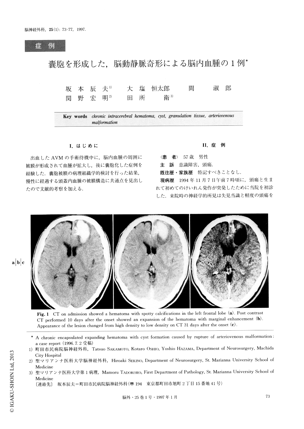
A 57-year-old male was admitted to our hospital complaining of a headache with disturbance of con-sciousness on November 7, 1994. CT scans revealed an intracerebral hematoma of 25ml in volume in the left frontal lobe with adjacent spotty calcification. An arte-riovenous malformation (AVM) with a nidus of 2.0×2.0cm in size was found to be the cause of the hemato-ma by cerebral angiography. Since the patient com-plained of mild right hemiparesis 10 days after the onset, CT scans were taken and a slightly enlarged hematoma with capsule formation was observed. On the 31st day after the onset, the hematoma had li-quefied and the surrounding capsule was clearly visible on CT. Since the patient's only symptom was a slight headache, and he displayed no other serious conditions, a palliative operation was planned. The AVM was re-moved and the capsule was resected 41 days after the onset.
The capsule or cyst wall of the liquefied hematoma was composed of three layers: a granulation layer with a neovascular system on the inside, a collagenous layer in the middle, and a reactive brain tissue layer on the outside. The structure of the capsule was the same as the structure of the cyst wall in chronic hematomas that have been reported as cystic AVM or encapsulated expanding hematoma in the literature. We would there-fore like to propose that chronic encapsulated expand-ing hematomas form with time due to intermittent bleeding or exudation form the neovascular system of a cyst wall.
Copyright © 1997, Igaku-Shoin Ltd. All rights reserved.