Japanese
English
- 有料閲覧
PPV購入案内
1,320円 (1,200円+税10%) こちらは、969ページから973ページの文献です。購入には医書.jp本会員登録が必要です。
会員登録完了後に改めて上記「購入サイトへ」を選択してください。
または「購入サイトへ」選択後に「購入ログイン」、「新規会員登録」の順に進んでください。
会員登録について詳しくはをご確認ください。
- Abstract 文献概要
- 1ページ目 Look Inside
- 参考文献 Reference
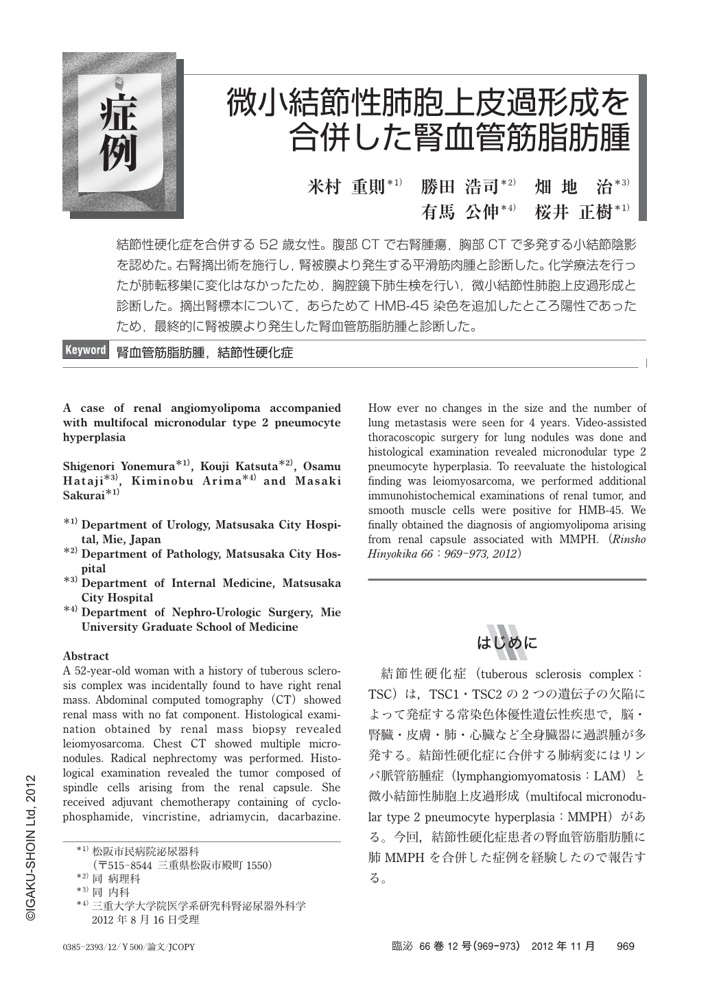
結節性硬化症を合併する52歳女性。腹部CTで右腎腫瘍,胸部CTで多発する小結節陰影を認めた。右腎摘出術を施行し,腎被膜より発生する平滑筋肉腫と診断した。化学療法を行ったが肺転移巣に変化はなかったため,胸腔鏡下肺生検を行い,微小結節性肺胞上皮過形成と診断した。摘出腎標本について,あらためてHMB-45染色を追加したところ陽性であったため,最終的に腎被膜より発生した腎血管筋脂肪腫と診断した。
A 52-year-old woman with a history of tuberous sclerosis complex was incidentally found to have right renal mass. Abdominal computed tomography(CT)showed renal mass with no fat component. Histological examination obtained by renal mass biopsy revealed leiomyosarcoma. Chest CT showed multiple micronodules. Radical nephrectomy was performed. Histological examination revealed the tumor composed of spindle cells arising from the renal capsule. She received adjuvant chemotherapy containing of cyclophosphamide, vincristine, adriamycin, dacarbazine. How ever no changes in the size and the number of lung metastasis were seen for 4 years. Video-assisted thoracoscopic surgery for lung nodules was done and histological examination revealed micronodular type 2 pneumocyte hyperplasia. To reevaluate the histological finding was leiomyosarcoma, we performed additional immunohistochemical examinations of renal tumor, and smooth muscle cells were positive for HMB-45. We finally obtained the diagnosis of angiomyolipoma arising from renal capsule associated with MMPH.
Copyright © 2012, Igaku-Shoin Ltd. All rights reserved.