Japanese
English
- 有料閲覧
PPV購入案内
1,320円 (1,200円+税10%) こちらは、165ページから167ページの文献です。購入には医書.jp本会員登録が必要です。
会員登録完了後に改めて上記「購入サイトへ」を選択してください。
または「購入サイトへ」選択後に「購入ログイン」、「新規会員登録」の順に進んでください。
会員登録について詳しくはをご確認ください。
- Abstract 文献概要
- 1ページ目 Look Inside
- 参考文献 Reference
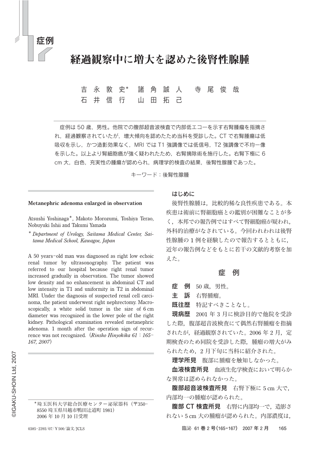
症例は50歳,男性。他院での腹部超音波検査で内部低エコーを示す右腎腫瘤を指摘され,経過観察されていたが,増大傾向を認めたため当科を受診した。CTで右腎腫瘍は低吸収を示し,かつ造影効果なく,MRIではT1強調像では低信号,T2強調像で不均一像を示した。以上より腎細胞癌が強く疑われたため,右腎摘除術を施行した。右腎下極に6cm大,白色,充実性の腫瘍が認められ,病理学的検査の結果,後腎性腺腫であった。
A 50 years-old man was diagnosed as right low echoic renal tumor by ultrasonography. The patient was referred to our hospital because right renal tumor increased gradually in observation. The tumor showed low density and no enhancement in abdominal CT and low intensity in T1 and uniformity in T2 in abdominal MRI. Under the diagnosis of suspected renal cell carcinoma, the patient underwent right nephrectomy. Macroscopically, a white solid tumor in the size of 6cm diameter was recognized in the lower pole of the right kidney. Pathological examination revealed metanephric adenoma. 1 month after the operation sign of recurrence was not recognized.(Rinsho Hinyokika 61:165-167, 2007)
Copyright © 2007, Igaku-Shoin Ltd. All rights reserved.