Japanese
English
- 有料閲覧
PPV購入案内
1,320円 (1,200円+税10%) こちらは、211ページから216ページの文献です。購入には医書.jp本会員登録が必要です。
会員登録完了後に改めて上記「購入サイトへ」を選択してください。
または「購入サイトへ」選択後に「購入ログイン」、「新規会員登録」の順に進んでください。
会員登録について詳しくはをご確認ください。
- Abstract 文献概要
- 1ページ目 Look Inside
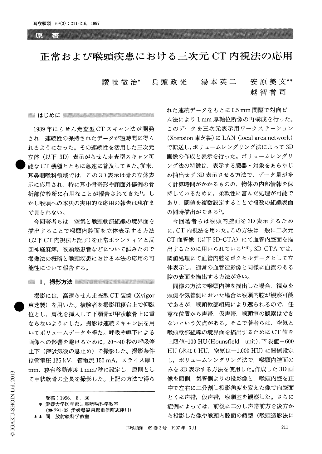
はじめに
1989年にらせん走査型CTスキャン法が開発され,連続性の保持されたデータが短時間に得られるようになった。その連続性を活用した三次元立体(以下3D)表示がらせん走査型スキャン可能なCT機種とともに急速に普及してきた。従来,耳鼻咽喉科領域では,この3D表示は骨の立体表示に応用され,特に耳小骨奇形や顔面外傷例の骨折部位診断に有用なことが報告されてきた1)。しかし喉頭への本法の実用的な応用の報告は現在まで見られない。
今回著者らは,空気と喉頭軟部組織の境界面を描出することで喉頭内腔面を立体表示する方法(以下CT内視法と記す)を正常ボランティアと反回神経麻痺,喉頭癌患者などについて試みたので撮像法の概略と喉頭疾患における本法の応用の可能性について報告する。
The recent development of helical (spiral) computed tomography allows collection of volumetric data to obtain high quality three-dimen-sional (3D) reconstructed images. The authors applied the 3D CT endoscopic imaging technique to assess normal and pathologic laryngeal structures. The latter included trauma, vocal fold atrophy, cancer of the larynx and recurrent nerve palsy. This technique was able to show normal laryngeal struc-tures and characteristic findings of each pathology. The 3D CT endoscopic images can be rotated around any axis, allowing optimal depiction of pathologic lesion. The use of 3D CT endoscopic technique provides the display of the location and extent of pathology and affords accurate therapeu-tic planning.
Copyright © 1997, Igaku-Shoin Ltd. All rights reserved.