Japanese
English
- 有料閲覧
PPV購入案内
1,320円 (1,200円+税10%) こちらは、627ページから632ページの文献です。購入には医書.jp本会員登録が必要です。
会員登録完了後に改めて上記「購入サイトへ」を選択してください。
または「購入サイトへ」選択後に「購入ログイン」、「新規会員登録」の順に進んでください。
会員登録について詳しくはをご確認ください。
- Abstract 文献概要
- 1ページ目 Look Inside
I.はじめに
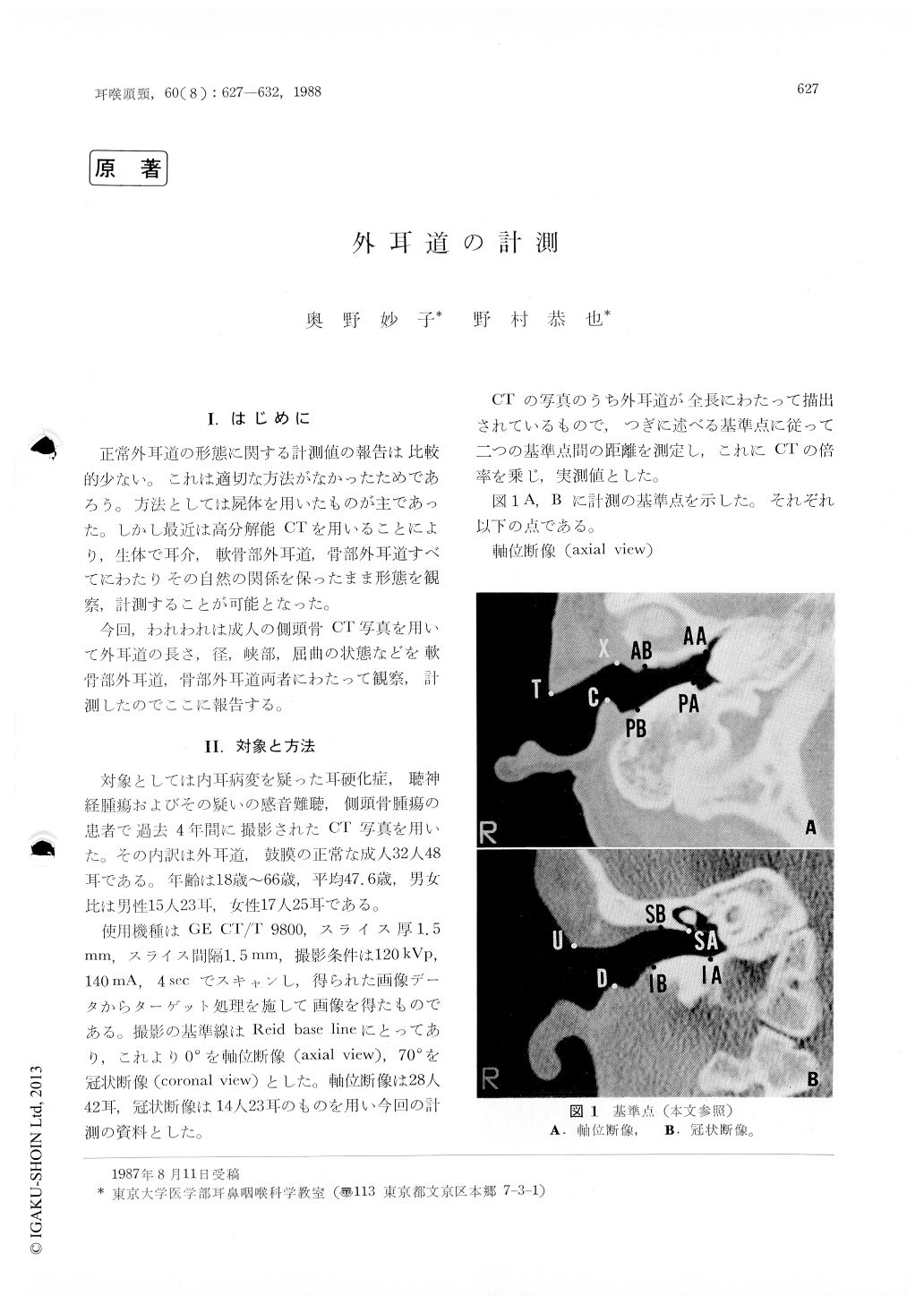
正常外耳道の形態に関する計測値の報告は比較的少ない。これは適切な方法がなかったためであろう。方法としては屍体を用いたものが主であった。しかし最近は高分解能CTを用いることにより,生体で耳介,軟骨部外耳道,骨部外耳道すべてにわたりその自然の関係を保ったまま形態を観察,計測することが可能となった。
今回,われわれは成人の側頭骨CT写真を用いて外耳道の長さ,径,峡部,屈曲の状態などを軟骨部外耳道,骨部外耳道両者こわたって観察,計測したのでここに報告する。
The length, width and angle of the normal human external auditory canal were measured using computed tomography.
Total length of the canal was measured : an-terior wall 34.5 mm, posterior wall 23.8 mm, superior wall 25.7 mm and inferior wall 30.0 mm. The proportion of the bony part/cartilagenous part was calculated: anterior 0.7, posterior 1, 2, superior 0.5 and inferior O.8.
The width of the canal was 10.4 mm × 12.4mm at the external orifice, 8.7 mm × 8.8 mm at the outer edge of the bony part and 9.1 mm × 9.1 mm at the annulus of the drum. The isthmus of the canal was located at the center of the bony canal anteroinferiorly and at the annulus of the drum posterosuperiorly. The width of the canal was 4.5 mm × 4.8 mm at the isthmus.
Copyright © 1988, Igaku-Shoin Ltd. All rights reserved.