Japanese
English
- 有料閲覧
PPV購入案内
1,320円 (1,200円+税10%) こちらは、617ページから620ページの文献です。購入には医書.jp本会員登録が必要です。
会員登録完了後に改めて上記「購入サイトへ」を選択してください。
または「購入サイトへ」選択後に「購入ログイン」、「新規会員登録」の順に進んでください。
会員登録について詳しくはをご確認ください。
- Abstract 文献概要
- 1ページ目 Look Inside
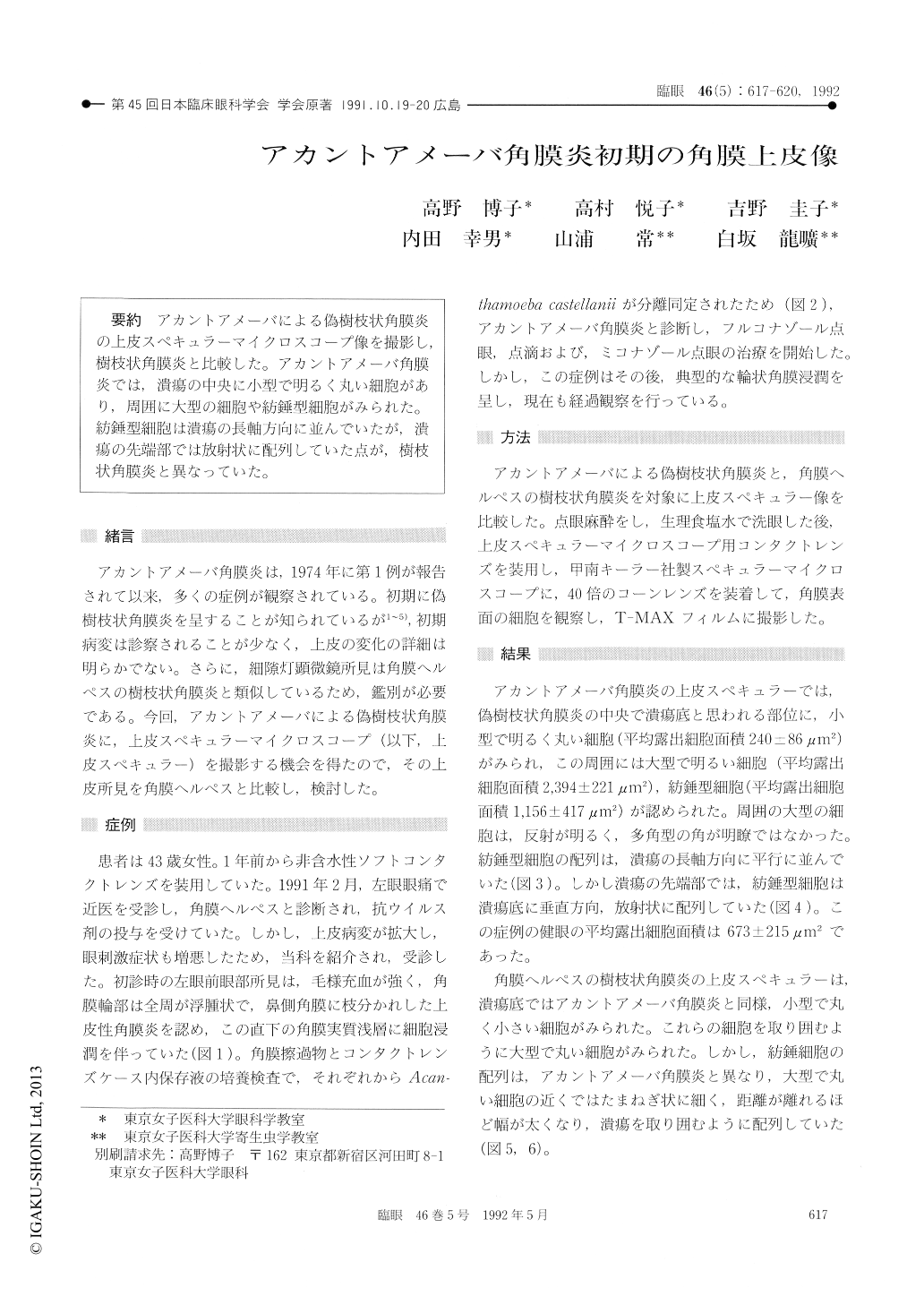
アカントアメーバによる偽樹枝状角膜炎の上皮スペキュラーマイクロスコープ像を撮影し,樹枝状角膜炎と比較した。アカントアメーバ角膜炎では,潰瘍の中央に小型で明るく丸い細胞があり,周囲に大型の細胞や紡錘型細胞がみられた。紡錘型細胞は潰瘍の長軸方向に並んでいたが,潰瘍の先端部では放射状に配列していた点が,樹枝状角膜炎と異なっていた。
A 43-year-old woman presented with pseudoden-dritic keratitis caused by Acanthamoeba castellanii. She had been wearing soft contact lens for the past one year. By specular microscopy, we observedsmall, round, bright cells in the center of the pseudodendritic lesion with similar appearance as dendritic keratitis. Additionally, there were large cells and spindle-shaped cells around the small ones. Spindle-shaped cells were arranged parallel to the direction of pseudendritic keratitis. As unique feature differentiating them from dendritic keratitis, they were arranged radially at the tip of the dendritic lesions.
Copyright © 1992, Igaku-Shoin Ltd. All rights reserved.