Japanese
English
- 有料閲覧
PPV購入案内
1,320円 (1,200円+税10%) こちらは、469ページから472ページの文献です。購入には医書.jp本会員登録が必要です。
会員登録完了後に改めて上記「購入サイトへ」を選択してください。
または「購入サイトへ」選択後に「購入ログイン」、「新規会員登録」の順に進んでください。
会員登録について詳しくはをご確認ください。
- Abstract 文献概要
- 1ページ目 Look Inside
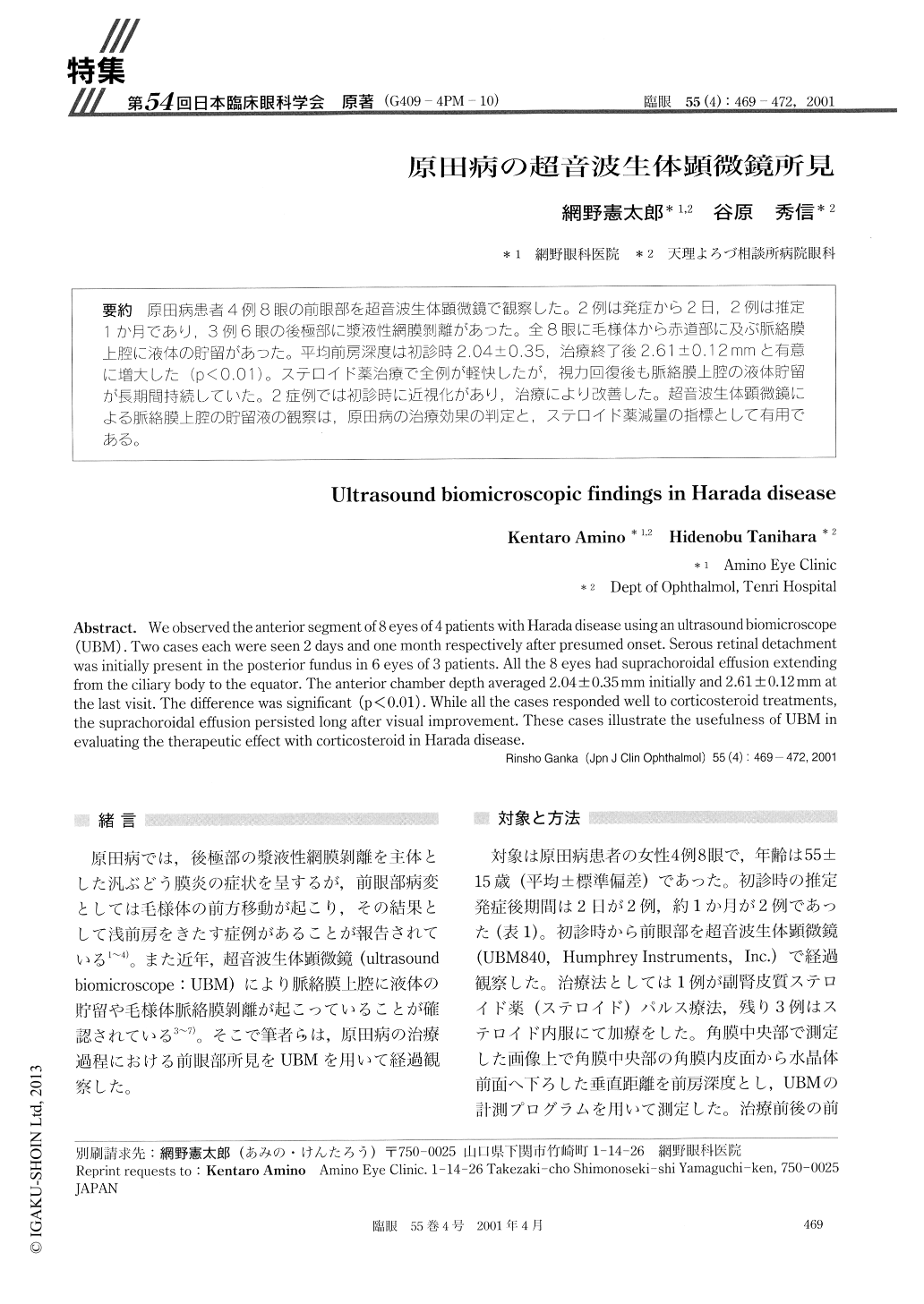
原田病患者4例8眼の前眼部を超音波生体顕微鏡で観察した。2例は発症から2日,2例は推定1か月であり,3例6眼の後極部に漿液性網膜剥離があった。全8眼に毛様体から赤道部に及ぶ脈絡膜上腔に液体の貯留があった。平均前房深度は初診時2.04±0.35,治療終了後2.61±0.12mmと有意に増大した(p<0.01)。ステロイド薬治療で全例が軽快したが,視力回復後も脈絡膜上腔の液体貯留が長期間持続していた。2症例では初診時に近視化があり,治療により改善した。超音波生体顕微鏡による脈絡膜上腔の貯留液の観察は,原田病の治療効果の判定と,ステロイド薬減量の指標として有用である。
We observed the anterior segment of 8 eyes of 4 patients with Harada disease using an ultrasound biomicroscope (UBM). Two cases each were seen 2 days and one month respectively after presumed onset. Serous retinal detachment was initially present in the posterior fundus in 6 eyes of 3 patients. All the 8 eyes had suprachoroidal effusion extending from the ciliary body to the equator. The anterior chamber depth averaged 2.04±0.35mm initially and 2.61±0.12mm at the last visit. The difference was significant (p<0.01). While all the cases responded well to corticosteroid treatments, the suprachoroidal effusion persisted long after visual improvement. These cases illustrate the usefulness of UBM in evaluating the therapeutic effect with corticosteroid in Harada disease.
Copyright © 2001, Igaku-Shoin Ltd. All rights reserved.