Japanese
English
- 有料閲覧
PPV購入案内
1,320円 (1,200円+税10%) こちらは、621ページから624ページの文献です。購入には医書.jp本会員登録が必要です。
会員登録完了後に改めて上記「購入サイトへ」を選択してください。
または「購入サイトへ」選択後に「購入ログイン」、「新規会員登録」の順に進んでください。
会員登録について詳しくはをご確認ください。
- Abstract 文献概要
- 1ページ目 Look Inside
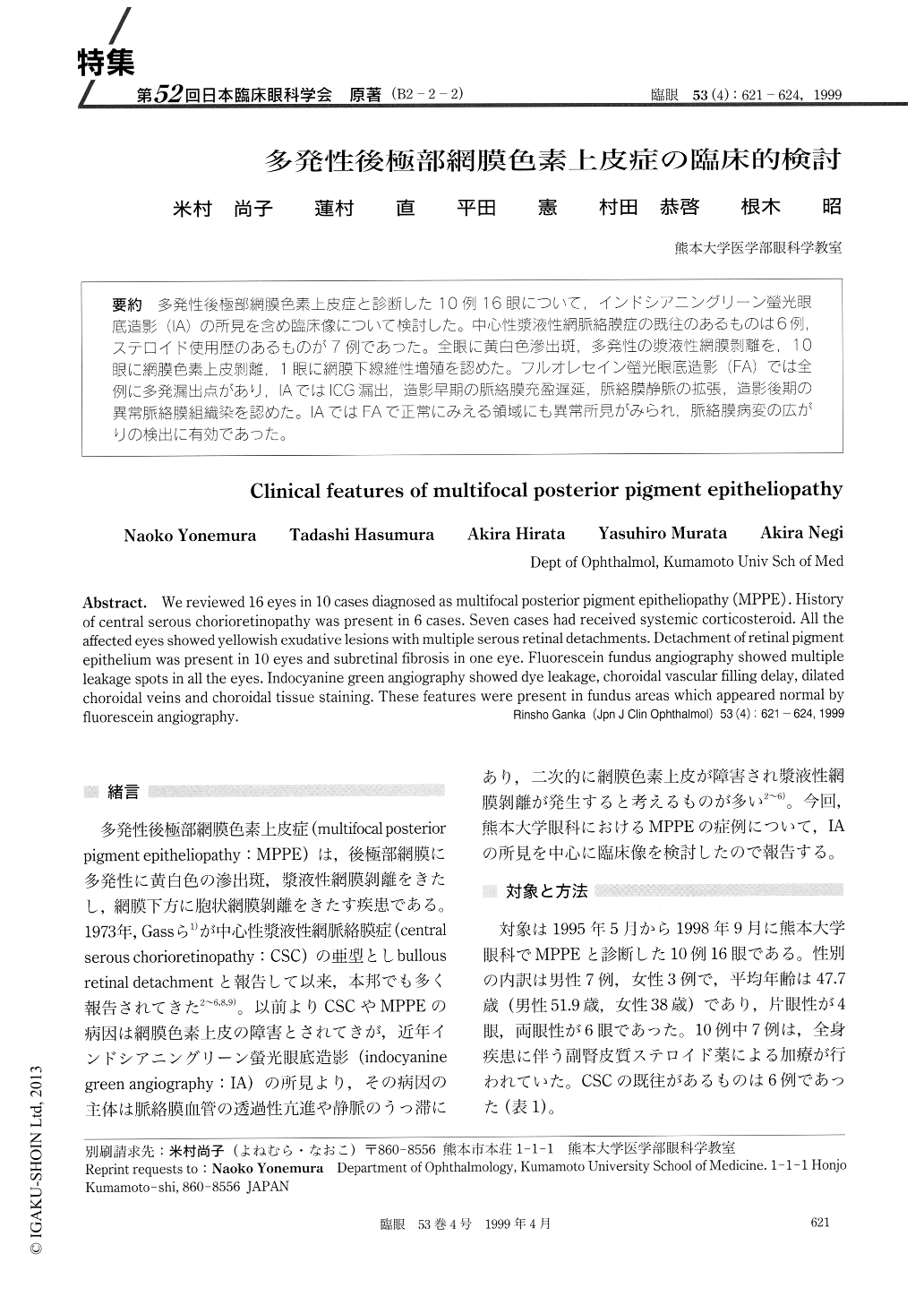
(B2-2-2) 多発性後極部網膜色素上皮症と診断した10例、6眼について,インドシアニングリーン螢光眼底造影(IA)の所見を含め臨床像について検討した。中心性漿液性網脈絡膜症の既往のあるものは6例,ステロイド使用歴のあるものが7例であった。全眼に黄白色滲出斑,多発性の漿液性網膜剥離を,10眼に網膜色素上皮剥離.1眼に網膜下線維性増殖を認めた。フルオレセイン螢光眼底造影(FA)では全例に多発漏出点があり,IAではlCG漏出,造影早期の脈絡膜充盈遅延,脈絡膜静脈の拡張,造影後期の異常脈絡膜組織染を認めた。IAではFAで正常にみえる領域にも異常所見がみられ,脈絡膜病変の広がりの検出に有効であった。
We reviewed 16 eyes in 10 cases diagnosed as multifocal posterior pigment epitheliopathy (MPPE) . History of central serous chorioretinopathy was present in 6 cases. Seven cases had received systemic corticosteroid. All the affected eyes showed yellowish exudative lesions with multiple serous retinal detachments. Detachment of retinal pigment epithelium was present in 10 eyes and subretinal fibrosis in one eye. Fluorescein fundus angiography showed multiple leakage spots in all the eyes. Indocyanine green angiography showed dye leakage, choroidal vascular filling delay, dilated choroidal veins and choroidal tissue staining. These features were present in fundus areas which appeared normal by fluorescein angiography.
Copyright © 1999, Igaku-Shoin Ltd. All rights reserved.