Japanese
English
- 有料閲覧
PPV購入案内
1,320円 (1,200円+税10%) こちらは、277ページから280ページの文献です。購入には医書.jp本会員登録が必要です。
会員登録完了後に改めて上記「購入サイトへ」を選択してください。
または「購入サイトへ」選択後に「購入ログイン」、「新規会員登録」の順に進んでください。
会員登録について詳しくはをご確認ください。
- Abstract 文献概要
- 1ページ目 Look Inside
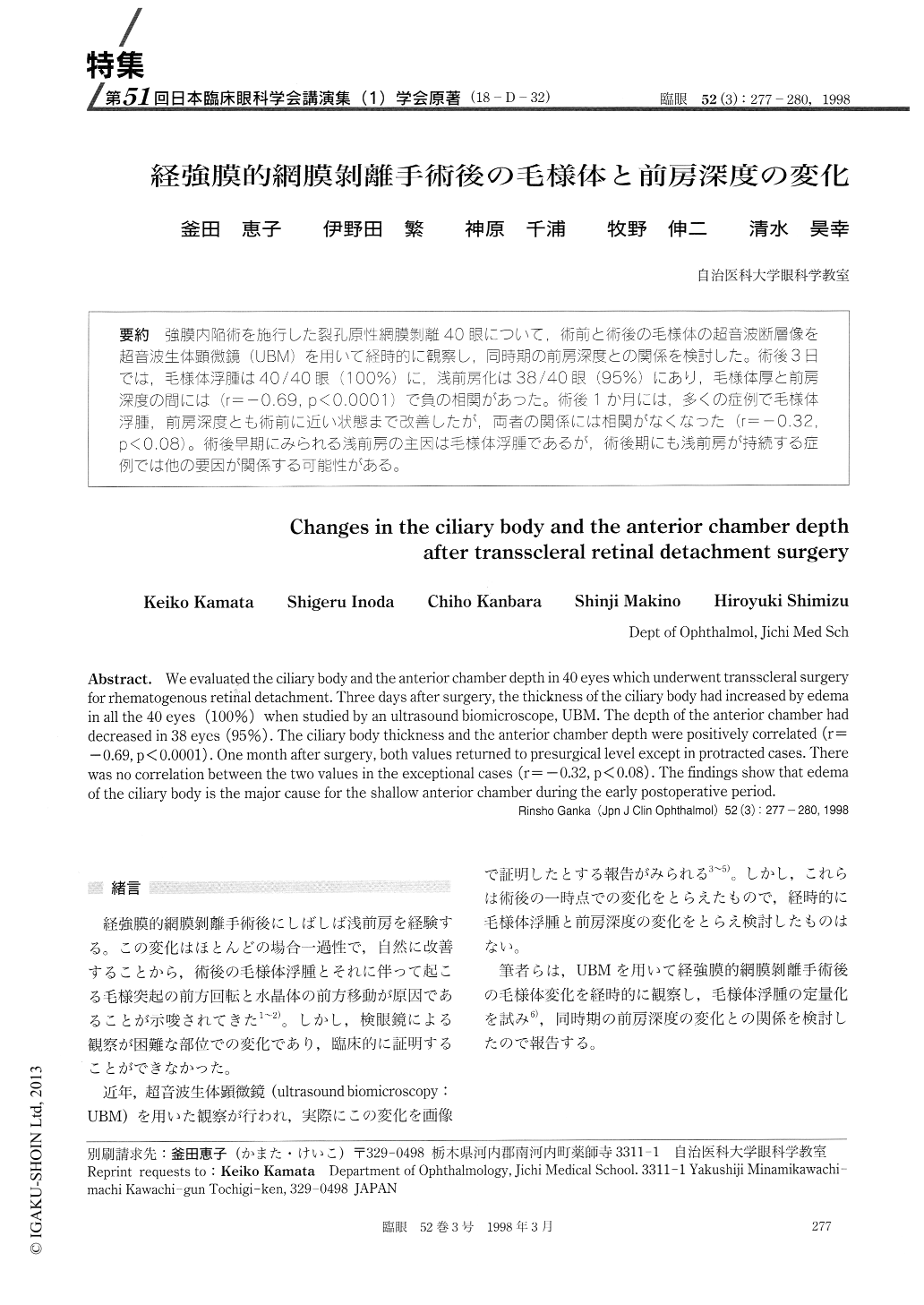
(18-D-32) 強膜内陥術を施行した裂孔原性網膜剥離40眼について,術前と術後の毛様体の超音波断層像を超音波生体顕微鏡(UBM)を用いて経時的に観察し,同時期の前房深度との関係を検討した。術後3日では,毛様体浮腫は40/40眼(100%)に,浅前房化は38/40眼(95%)にあり,毛様体厚と前房深度の間には(r=−0.69,p<0.0001)で負の相関があった。術後1か月には,多くの症例で毛様体浮腫,前房深度とも術前に近い状態まで改善したが,両者の関係には相関がなくなった(r=−0.32,p<0.08)。術後早期にみられる浅前房の主因は毛様体浮腫であるが,術後期にも浅前房が持続する症例では他の要因が関係する可能性がある。
We evaluated the ciliary body and the anterior chamber depth in 40 eyes which underwent transscleral surgery for rhematogenous retinal detachment. Three days after surgery, the thickness of the ciliary body had increased by edema in all the 40 eyes (100%) when studied by an ultrasound biomicroscope, UBM. The depth of the anterior chamber had decreased in 38 eyes (95%). The ciliary body thickness and the anterior chamber depth were positively correlated (r=-0.69, p<0.0001) . One month after surgery, both values returned to presurgical level except in protracted cases. There was no correlation between the two values in the exceptional cases (r=-0.32, p<0.08). The findings show that edema of the ciliary body is the major cause for the shallow anterior chamber during the early postoperative period.
Copyright © 1998, Igaku-Shoin Ltd. All rights reserved.