Japanese
English
- 有料閲覧
PPV購入案内
1,320円 (1,200円+税10%) こちらは、1269ページから1272ページの文献です。購入には医書.jp本会員登録が必要です。
会員登録完了後に改めて上記「購入サイトへ」を選択してください。
または「購入サイトへ」選択後に「購入ログイン」、「新規会員登録」の順に進んでください。
会員登録について詳しくはをご確認ください。
- Abstract 文献概要
- 1ページ目 Look Inside
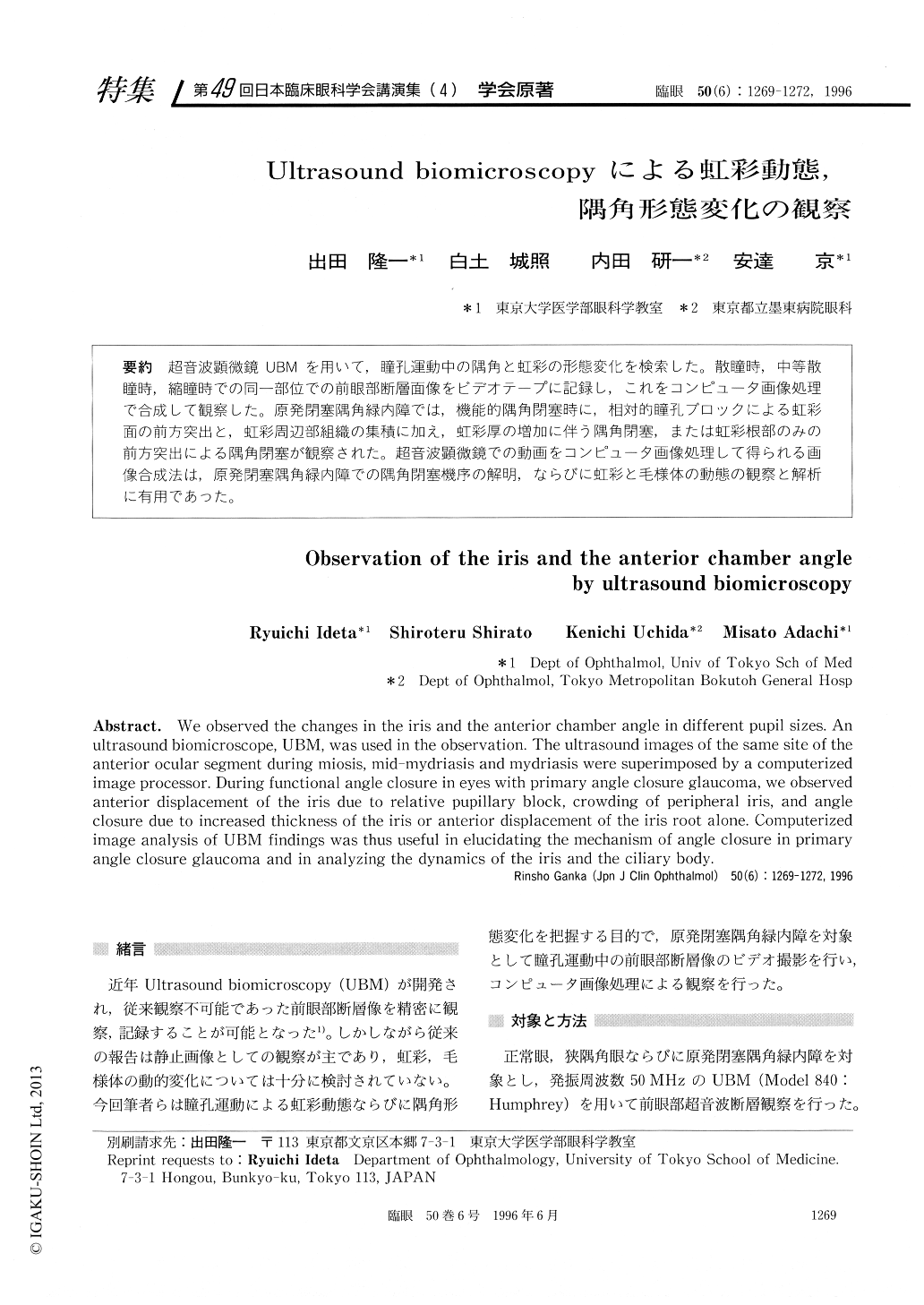
超音波顕微鏡UBMを用いて,瞳孔運動中の隅角と虹彩の形態変化を検索した。散瞳時,中等散瞳時,縮瞳時での同一部位での前眼部断層面像をビデオテープに記録し,これをコンピュータ画像処理で合成して観察した。原発閉塞隅角緑内障では,機能的隅角閉塞時に,相対的瞳孔ブロックによる虹彩面の前方突出と,虹彩周辺部組織の集積に加え,虹彩厚の増加に伴う隅角閉塞,または虹彩根部のみの前方突出による隅角閉塞が観察された。超音波顕微鏡での動画をコンピュータ画像処理して得られる画像合成法は,原発閉塞隅角緑内障での隅角閉塞機序の解明,ならびに虹彩と毛様体の動態の観察と解析に有用であった。
We observed the changes in the iris and the anterior chamber angle in different pupil sizes. An ultrasound biomicroscope, UBM, was used in the observation. The ultrasound images of the same site of the anterior ocular segment during miosis, mid-mydriasis and mydriasis were superimposed by a computerized image processor. During functional angle closure in eyes with primary angle closure glaucoma, we observed anterior displacement of the iris due to relative pupillary block, crowding of peripheral iris, and angle closure due to increased thickness of the iris or anterior displacement of the iris root alone. Computerized image analysis of UBM findings was thus useful in elucidating the mechanism of angle closure in primary angle closure glaucoma and in analyzing the dynamics of the iris and the ciliary body.
Copyright © 1996, Igaku-Shoin Ltd. All rights reserved.