Japanese
English
- 有料閲覧
PPV購入案内
1,320円 (1,200円+税10%) こちらは、1099ページから1104ページの文献です。購入には医書.jp本会員登録が必要です。
会員登録完了後に改めて上記「購入サイトへ」を選択してください。
または「購入サイトへ」選択後に「購入ログイン」、「新規会員登録」の順に進んでください。
会員登録について詳しくはをご確認ください。
- Abstract 文献概要
- 1ページ目 Look Inside
- 参考文献 Reference
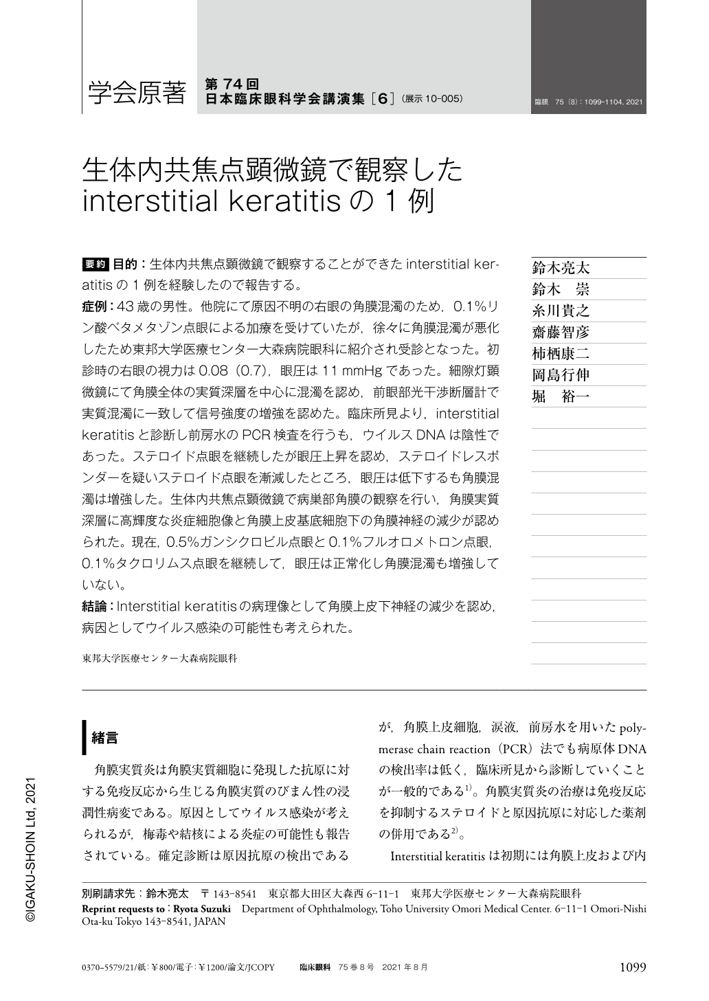
要約 目的:生体内共焦点顕微鏡で観察することができたinterstitial keratitisの1例を経験したので報告する。
症例:43歳の男性。他院にて原因不明の右眼の角膜混濁のため,0.1%リン酸ベタメタゾン点眼による加療を受けていたが,徐々に角膜混濁が悪化したため東邦大学医療センター大森病院眼科に紹介され受診となった。初診時の右眼の視力は0.08(0.7),眼圧は11mmHgであった。細隙灯顕微鏡にて角膜全体の実質深層を中心に混濁を認め,前眼部光干渉断層計で実質混濁に一致して信号強度の増強を認めた。臨床所見より,interstitial keratitisと診断し前房水のPCR検査を行うも,ウイルスDNAは陰性であった。ステロイド点眼を継続したが眼圧上昇を認め,ステロイドレスポンダーを疑いステロイド点眼を漸減したところ,眼圧は低下するも角膜混濁は増強した。生体内共焦点顕微鏡で病巣部角膜の観察を行い,角膜実質深層に高輝度な炎症細胞像と角膜上皮基底細胞下の角膜神経の減少が認められた。現在,0.5%ガンシクロビル点眼と0.1%フルオロメトロン点眼,0.1%タクロリムス点眼を継続して,眼圧は正常化し角膜混濁も増強していない。
結論:Interstitial keratitisの病理像として角膜上皮下神経の減少を認め,病因としてウイルス感染の可能性も考えられた。
Abstract Purpose:Here we report a case of interstitial keratitis using In vivo confocal microscopy.
Case:A 43-year-old man had been attending another hospital for an unexplained corneal opacity in his right eye of unknown origin. He was treated with 0.1% betamethasone sodium phosphate eye drops, but his corneal opacity gradually worsened and he was referred to our department. The patient's right eye visual acuity was 0.08(0.7)and intraocular pressure was 11 mmHg on the initial examination. Slit-lamp microscopy showed opacity centered in the deep parenchyma of the entire cornea, while anterior optical coherence tomography showed enhanced signal intensity, consistent with parenchymal opacity. Based on the clinical findings, interstitial keratitis was diagnosed and polymerase chain reaction testing of the aqueous humor was negative for viral DNA. The 0.1% betamethasone sodium phosphate eye drops were continued, but his intraocular pressure increased to over 30 mmHg which suggested that he was a steroid responder. When the 0.1% betamethasone sodium phosphate eye drops were tapered off, his intraocular pressure decreased but his corneal opacity increased. In vivo confocal microscopy of the diseased cornea revealed hyperintense inflammatory cells in the deep corneal parenchyma and loss of the corneal nerve under the basal corneal epithelial cells. Subsequently, 0.1% ganciclovir eye drops, 0.1% fluorometholone eye drops, and 0.1% tacrolimus eye drops were prescribed, which normalized the intraocular pressure and did not enhance the corneal opacity.
Conclusion:The pathology of interstitial keratitis showed corneal nerve loss, and viral infection was considered as a possible etiology.
Copyright © 2021, Igaku-Shoin Ltd. All rights reserved.