Japanese
English
特集 第42回日本臨床眼科学会講演集(2)1988年9月 東京
学会原著
中心性漿液性脈絡網膜症にみられる黄斑部網膜色素上皮の顆粒状萎縮斑
Granular atrophic fleck of retinal pigment epithelium in central serous chorioretinopathy
吉岡 久春
1
,
津村 知子
1
,
石橋 達朗
2
Hisaharu Yoshioka
1
,
Tomoko Tsumura
1
,
Tatsuro Ishibashi
2
1久留米大学医学部眼科
2九州大学医学部眼科
1Dept of Ophthalmol, Kurume Univ Sch of Med
2Dept of Ophthalmol, Fac of Med, Kyusyu Univ
pp.363-366
発行日 1989年3月15日
Published Date 1989/3/15
DOI https://doi.org/10.11477/mf.1410210669
- 有料閲覧
PPV購入案内
1,320円 (1,200円+税10%) こちらは、363ページから366ページの文献です。
PPV(ペイ・パー・ビュー)とは、論文(記事)単位で購入・閲覧ができるサービスです。
購入には医書.jp本会員登録が必要です。
会員登録完了後に改めて上記「購入サイトへ」を選択してください。
または「購入サイトへ」選択後に「購入ログイン」、「新規会員登録」の順に進んでください。
会員登録について詳しくはをご確認ください。
購入には医書.jp本会員登録が必要です。
会員登録完了後に改めて上記「購入サイトへ」を選択してください。
または「購入サイトへ」選択後に「購入ログイン」、「新規会員登録」の順に進んでください。
会員登録について詳しくはをご確認ください。
- Abstract 文献概要
- 1ページ目 Look Inside
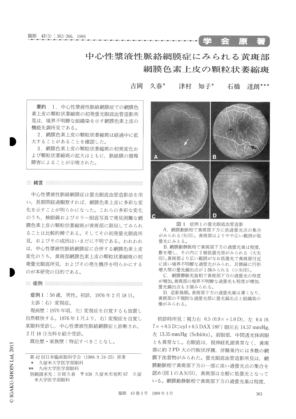
1.中心性漿液性脈絡網膜症での網膜色素上皮の顆粒状萎縮斑の初発螢光眼底血管造影所見は,境界不明瞭な組織染を示す網膜色素上皮の機能失調所見である。
2.網膜色素上皮の顆粒状萎縮斑は経過中に拡大することがあることを確認した。
3.網膜色素土皮の顆粒状萎縮斑の初発変化および顆粒状萎縮斑の拡大はともに,脈絡膜の循環障害によることが示唆された。
We observed two cases with central serous chor-ioretinopathy by means of fluorescein fundus angio-graphy. Both were males aged 39 and 50 years each. During the early phase in the angiogram, we detect-ed granular atrophic fleck in the macula. The fleck was located at the level of retinal pigment epith-elium and later turned into ill-defined patch due to tissue staining. The fleck increased in size during the course of the disease. Choroidal circulatory disorder seemed to underlie the observed atrophic fleck and its enlargement.
Copyright © 1989, Igaku-Shoin Ltd. All rights reserved.